01 / Morfologi

Ciri dan struktur

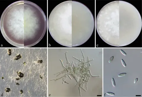
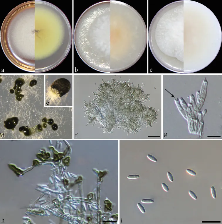
Colonies on PDA, CMA and OA with white aerial mycelium, approx. 6–7 cm diam. after 7 d at 25 °C, giving rise to dark green or blank sporodochia scattered or gregarious on the surface, covered by olivaceous green pillars of conidia, reverse on PDA sienna. Hyphae hyaline, smooth, branched, 1–2 μm wide. Conidiomata synnematous, solitary, 60–250 μm high, 30–80 μm wide at the base, 60–150 μm at the apex, with setose hyphae surrounding a green agglutinated mass of conidia. Stroma well developed, hyaline, of textura angularis. Setae absent. Conidiophores arising from the basal stroma, branched, initially hyaline and becoming pigmented and verrucose with age covered by an olivaceous green mucoid layer, up to 30 µm long. Conidiogenous cell phialidic, clavate to cylindrical, hyaline, smooth, 5–10 × 1–2 µm, becoming pigmented and verrucose with age, with conspicuous collarettes and periclinal thickenings. Conidia aseptate, smooth, hyaline, ellipsoidal to fusiform, 6–8 × 2–3 µm (av. 7 ± 0.4× 2 ± 0.2 µm, n = 50).

Sebagian uraian ditampilkan dalam bahasa sumber; terjemahannya belum tersedia atau belum lolos pemeriksaan.

Rujukan: [1]

Pengukuran mengikuti objek, spesimen atau kultur, dan metode yang disebutkan sumber. Baca uraian asli untuk konteks lengkap.

02 / Kehidupan

Habitat dan peran ekologis

China, Hainan Province, Haikou, isolated from leaves of Imperata cylindrica, 10 Mar 2018, J.M. Liang and L. Cai, holotype HMAS 247953, ex-holotype culture CGMCC3.19198 = LC12140. Colonies on PDA, CMA and OA with white aerial mycelium, approx. 6–7 cm diam. after 7 d at 25 °C, giving rise to dark green or blank sporodochia scattered or gregarious on the surface, covered by olivaceous green pillars of conidia, reverse on PDA sienna. Hyphae hyaline, smooth, branched, 1–2 μm wide. Conidiomata synnematous, solitary, 60–250 μm high, 30–80 μm wide at the base, 60–150 μm at the apex, with setose hyphae surrounding a green agglutinated mass of conidia. Stroma well developed, hyaline, of textura angularis. Setae absent. Conidiophores arising from the basal stroma, branched, initially hyaline and becoming pigmented and verrucose with age covered by an olivaceous green mucoid layer, up to 30 µm long. Conidiogenous cell phialidic, clavate to cylindrical, hyaline, smooth, 5–10 × 1–2 µm, becoming pigmented and verrucose with age, with conspicuous collarettes and periclinal thickenings. Conidia aseptate, smooth, hyaline, ellipsoidal to fusiform, 6–8 × 2–3 µm (av. 7 ± 0.4× 2 ± 0.2 µm, n = 50). Alfaria poae formed a well-supported clade in Alfaria (Fig. 1).

Sebagian uraian ditampilkan dalam bahasa sumber; terjemahannya belum tersedia atau belum lolos pemeriksaan.

Rujukan: [1]

Habitat atau konteks temuan
kultur yang didokumentasikan
Substrat
Informasi belum tersedia pada sumber rilis ini.
Peran atau konteks
Belum dikategorikan pada rilis ini.
Temukan hubungan serupa
03 / Siklus hidup

Reproduksi

Colonies on PDA, CMA and OA with white aerial mycelium, approx. 6–7 cm diam. after 7 d at 25 °C, giving rise to dark green or blank sporodochia scattered or gregarious on the surface, covered by olivaceous green pillars of conidia, reverse on PDA sienna. Hyphae hyaline, smooth, branched, 1–2 μm wide. Conidiomata synnematous, solitary, 60–250 μm high, 30–80 μm wide at the base, 60–150 μm at the apex, with setose hyphae surrounding a green agglutinated mass of conidia. Stroma well developed, hyaline, of textura angularis. Setae absent. Conidiophores arising from the basal stroma, branched, initially hyaline and becoming pigmented and verrucose with age covered by an olivaceous green mucoid layer, up to 30 µm long.

Sebagian uraian ditampilkan dalam bahasa sumber; terjemahannya belum tersedia atau belum lolos pemeriksaan.

Rujukan: [1]

04 / Batas informasi

Catatan identitas dan cakupan

  • Nama diterima mengikuti Catalogue of Life Base2026-09-11; uraian mempertahankan konsep dan nama sumber aslinya.
  • Preserve original treatment localities, hosts and substrate scope. Materials examined describe documented specimens, not the full global range.
  • Impor dan terjemahan otomatis belum diperiksa editorial per profil. Struktur reproduktif yang disebutkan tidak membuktikan seluruh siklus hidup diketahui.
  • Pengukuran pada uraian mengikuti spesimen, kultur, dan metode sumber; angka tidak diperluas ke semua populasi.

Catatan distribusi dalam narasi mengikuti sumbernya. Dokumentasi spesimen atau foto mengikuti catatan sumber. Bukti wilayah Indonesia dan penilaian konservasi tidak disimpulkan dari gambar.

05 / Dokumentasi

Melihat dengan konteks

Alfaria poae (from ex-type strain CGMCC3.19198) a–c colony on PDA, CMA, OA d–e conidiomata on SNA f synnematous conidioma g conidiogenous cells, the arrow showing conspicuous collarette h aged conidiophores i conidia. Scale bars: 50 μm (f); 5 μm (g); 10 μm (h, i).

Jenis
Pelat ilmiah
Kredit
Junmin Liang, Guangshuo Li, Shiyue Zhou, Meiqi Zhao, Lei Cai
Lisensi
CC BY 4.0
Sumber media
Buka entri sumber

Gambar menunjukkan objek menurut identifikasi sumber, bukan verifikasi independen spesimen oleh Fungi. Tidak ada skala baru yang ditambahkan.

06 / Rujukan

Sumber profil

  1. [1] Myrothecium-like new species from turfgrasses and associated rhizosphere — Alfaria poaeBagian sumber: PMC6583245_section_31
    Tanggal penghimpunan sumber: 2026-10-12
    Teks sumber: CC BY 4.0 · Liang J, Li G, Zhou S, Zhao M, Cai L.
  2. [2] Catalogue of Life Base2026-09-11 — Alfaria poaeBagian sumber: ColDP NameUsage.tsv ID BRKW; accepted species; source 2073
    Tanggal penghimpunan sumber: 2026-10-12
  3. [3] Dokumentasi visual — Alfaria poaeBagian sumber: Individual media record; identification follows the source, not independent determination
    Tanggal penghimpunan sumber: 2026-10-12
    Media sumber: CC BY 4.0 · Junmin Liang, Guangshuo Li, Shiyue Zhou, Meiqi Zhao, Lei Cai

Adaptasi teks: CC BY 4.0. Liang J, Li G, Zhou S, Zhao M, Cai L. — Myrothecium-like new species from turfgrasses and associated rhizosphere — Alfaria poae

Uraian asli dan riwayat sumber

Riwayat atau halaman sumber

Ringkasan sumber

Colonies on PDA, CMA and OA with white aerial mycelium, approx. 6–7 cm diam. after 7 d at 25 °C, giving rise to dark green or blank sporodochia scattered or gregarious on the surface, covered by olivaceous green pillars of conidia, reverse on PDA sienna. Hyphae hyaline, smooth, branched, 1–2 μm wide. Conidiomata synnematous, solitary, 60–250 μm high, 30–80 μm wide at the base, 60–150 μm at the apex, with setose hyphae surrounding a green agglutinated mass of conidia. Stroma well developed, hyaline, of textura angularis.

Revisi sumber

Morfologi sumber

Colonies on PDA, CMA and OA with white aerial mycelium, approx. 6–7 cm diam. after 7 d at 25 °C, giving rise to dark green or blank sporodochia scattered or gregarious on the surface, covered by olivaceous green pillars of conidia, reverse on PDA sienna. Hyphae hyaline, smooth, branched, 1–2 μm wide. Conidiomata synnematous, solitary, 60–250 μm high, 30–80 μm wide at the base, 60–150 μm at the apex, with setose hyphae surrounding a green agglutinated mass of conidia. Stroma well developed, hyaline, of textura angularis. Setae absent. Conidiophores arising from the basal stroma, branched, initially hyaline and becoming pigmented and verrucose with age covered by an olivaceous green mucoid layer, up to 30 µm long. Conidiogenous cell phialidic, clavate to cylindrical, hyaline, smooth, 5–10 × 1–2 µm, becoming pigmented and verrucose with age, with conspicuous collarettes and periclinal thickenings. Conidia aseptate, smooth, hyaline, ellipsoidal to fusiform, 6–8 × 2–3 µm (av. 7 ± 0.4× 2 ± 0.2 µm, n = 50).

Revisi sumber

Ekologi sumber

China, Hainan Province, Haikou, isolated from leaves of Imperata cylindrica, 10 Mar 2018, J.M. Liang and L. Cai, holotype HMAS 247953, ex-holotype culture CGMCC3.19198 = LC12140. Colonies on PDA, CMA and OA with white aerial mycelium, approx. 6–7 cm diam. after 7 d at 25 °C, giving rise to dark green or blank sporodochia scattered or gregarious on the surface, covered by olivaceous green pillars of conidia, reverse on PDA sienna. Hyphae hyaline, smooth, branched, 1–2 μm wide. Conidiomata synnematous, solitary, 60–250 μm high, 30–80 μm wide at the base, 60–150 μm at the apex, with setose hyphae surrounding a green agglutinated mass of conidia. Stroma well developed, hyaline, of textura angularis. Setae absent. Conidiophores arising from the basal stroma, branched, initially hyaline and becoming pigmented and verrucose with age covered by an olivaceous green mucoid layer, up to 30 µm long. Conidiogenous cell phialidic, clavate to cylindrical, hyaline, smooth, 5–10 × 1–2 µm, becoming pigmented and verrucose with age, with conspicuous collarettes and periclinal thickenings. Conidia aseptate, smooth, hyaline, ellipsoidal to fusiform, 6–8 × 2–3 µm (av. 7 ± 0.4× 2 ± 0.2 µm, n = 50). Alfaria poae formed a well-supported clade in Alfaria (Fig. 1).

Revisi sumber

Struktur reproduksi sumber

Colonies on PDA, CMA and OA with white aerial mycelium, approx. 6–7 cm diam. after 7 d at 25 °C, giving rise to dark green or blank sporodochia scattered or gregarious on the surface, covered by olivaceous green pillars of conidia, reverse on PDA sienna. Hyphae hyaline, smooth, branched, 1–2 μm wide. Conidiomata synnematous, solitary, 60–250 μm high, 30–80 μm wide at the base, 60–150 μm at the apex, with setose hyphae surrounding a green agglutinated mass of conidia. Stroma well developed, hyaline, of textura angularis. Setae absent. Conidiophores arising from the basal stroma, branched, initially hyaline and becoming pigmented and verrucose with age covered by an olivaceous green mucoid layer, up to 30 µm long.

Revisi sumber

Rujukan halaman ini

Fungi. Alfaria poae. Rilis data fungi-2026-10-12-expansion-2. https://fungi-atlas.mgadib.chatgpt.site/jamur/alfaria-poae/.

Status: impor sumber; pemeriksaan editorial per profil belum selesai. Untuk klaim biologis, gunakan rujukan pada bagian terkait dan periksa konteks sumber aslinya.