Ciri dan struktur
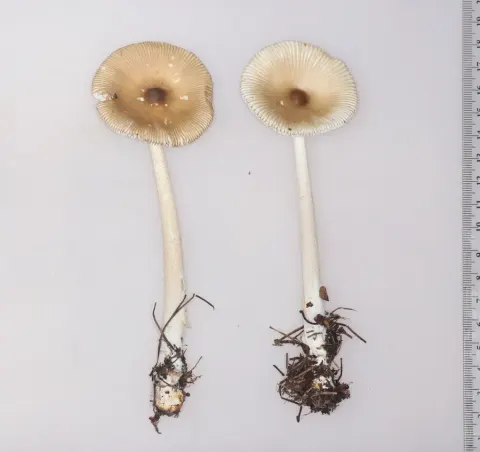
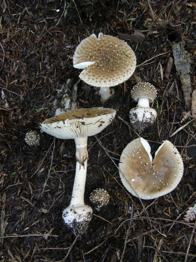
The shape of the A. australis cap is initially convex, later flattening out or even developing a central depression, and reaching diameters of 2-9 cm wide. The cap margin sometimes splits and rolls back to give a ragged appearance. The centre of the cap is dark buff, honey or isabelline, becoming paler to buff at the margin. The surface is sticky when young or wet, but dries out with age. The remnants of the volva form conical to pyramidal warts that are most densely aggregated in the center, but become sparse and low towards the margin. They are initially white then greyish-sepia or isabelline with white to buff tips. The gills are crowded closely together, free from attachment to the stem, 6 – 10 mm wide, and white. The lamellulae (short gills that do not extend fully from the cap edge to the stem) have truncated ends. The stem is 37 – 90 mm tall, 6 – 26 mm in diameter, and narrowest at center. It is hollow, and has an abruptly bulbous base that is between 14 and 38 mm in diameter. The surface of the stem above the level of the ring is white and covered in woolly tufts of mycelia; below the ring it is white with buff to greyish transverse, grooved bands. The base may or may not have a rim of volval remnants that are powdery, and a greyish-buff to greyish-sepia colour. The ring is membranous, white to buff, first hanging freely then later adhering to the stem. The flesh of the cap is white, occasionally pale isabelline under the center of the cap; the flesh of the stem is white. The spore print is white. The spores are typically 9–12 by 8–10.5 μm, spherical to ellipsoid, and thin-walled.
Habitat dan peran ekologis
A. australis hanya ditemukan di pulau-pulau utara dan selatan Selandia Baru, di mana tumbuh dalam hubungan mikoriza dengan bukit selatan (genus Nothofagus) (termasuk bukit baru Zealand red, bukit perak, bukit baru Zealand black, bukit keras), mānuka, dan kānuka. Jamur ini biasanya tumbuh sendiri, tetapi dalam kesempatan yang jarang telah ditemukan tumbuh dalam kelompok.
- Habitat atau konteks temuan
- Informasi belum tersedia pada sumber rilis ini.
- Substrat
- Informasi belum tersedia pada sumber rilis ini.
- Peran atau konteks
- Mitra tumbuhan
Reproduksi
The spore print is white. The spores are typically 9–12 by 8–10.5 μm, spherical to ellipsoid, and thin-walled. They are hyaline (translucent), and amyloid—meaning they will stain bluish-black to black in Melzer's reagent. The basidia are 43.5–76.5 by 10.5–17 μm, mostly four-spored, and clamped at their bases. There are abundant spherical, elliptic or club-shaped hyaline cells on the gill edges, measuring 16–39.5 by 10.5–27.5 μm. The cap cuticle is 220–270 μm wide, consisting of a gelatinised suprapellis (upper layer) and non-gelatinised subpellis (lower layer). The volval remnants on the cap consist of abundant spherical, club-shaped, or turnip-shaped cells, measuring 10–86 by 9–85 μm. These cells are umber in colour, and arranged in chains perpendicular to the cap surface, becoming smaller and paler at tip of the wart, subtended by moderately abundant hyphae that are 4–10 μm wide.
Catatan identitas dan cakupan
- Nama diterima mengikuti Catalogue of Life Base2026-09-11; uraian mempertahankan konsep dan nama sumber aslinya.
- Original geographic, specimen and life-cycle scope retained; source import awaits individual editorial review.
- Impor dan terjemahan otomatis belum diperiksa editorial per profil. Struktur reproduktif yang disebutkan tidak membuktikan seluruh siklus hidup diketahui.
- Pengukuran pada uraian mengikuti spesimen, kultur, dan metode sumber; angka tidak diperluas ke semua populasi.
Catatan distribusi dalam narasi mengikuti sumbernya. Dokumentasi spesimen atau foto mengikuti catatan sumber. Bukti wilayah Indonesia dan penilaian konservasi tidak disimpulkan dari gambar.
Melihat dengan konteks
Dokumentasi Amanita australis menurut asosiasi foto pada record takson iNaturalist. Kredit: David Whyte. Identitas mengikuti sumber; bukan penentuan spesimen independen.
- Jenis
- Fotografi
- Kredit
- (c) David Whyte, some rights reserved (CC BY-SA), uploaded by David Whyte
- Lisensi
- CC BY-SA 4.0
- Sumber media
- Buka entri sumber
Dokumentasi tambahan
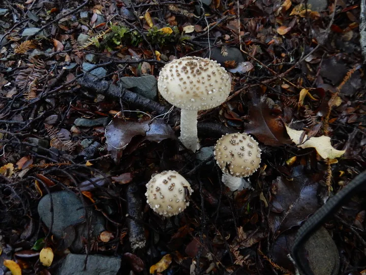
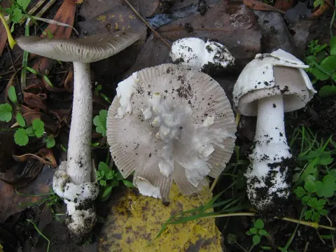
Foto yang diasosiasikan dengan Amanita australis pada iNaturalist. Identifikasi mengikuti sumber; bukan penentuan spesimen independen.
Kredit dan penggunaan
(c) Ray Jackson, some rights reserved (CC BY), uploaded by Ray Jackson
Ukuran dan format gambar disesuaikan tanpa pemotongan, retus, atau penambahan skala. Lisensi sumber dipertahankan.
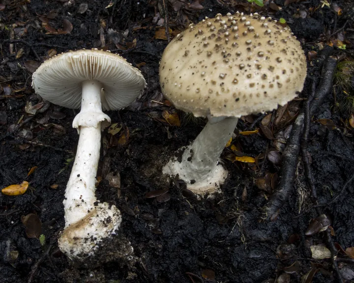
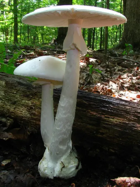
Foto yang diasosiasikan dengan Amanita australis pada iNaturalist. Identifikasi mengikuti sumber; bukan penentuan spesimen independen.
Kredit dan penggunaan
(c) Jerry Cooper, some rights reserved (CC BY), uploaded by Jerry Cooper
Ukuran dan format gambar disesuaikan tanpa pemotongan, retus, atau penambahan skala. Lisensi sumber dipertahankan.
Sumber profil
- [1] Amanita australis — Wikipedia (en)
- [2] Catalogue of Life Base2026-09-11 — Amanita australis
- [3] Dokumentasi visual — Amanita australis
- [4] Dokumentasi visual — Amanita australis
- [5] Dokumentasi visual — Amanita australis
Rujukan halaman ini
Fungi. Amanita australis. Rilis data fungi-2026-10-12-expansion-2. https://fungi-atlas.mgadib.chatgpt.site/jamur/amanita-australis/.
Status: impor sumber; pemeriksaan editorial per profil belum selesai. Untuk klaim biologis, gunakan rujukan pada bagian terkait dan periksa konteks sumber aslinya.