01 / Morfologi

Ciri dan struktur

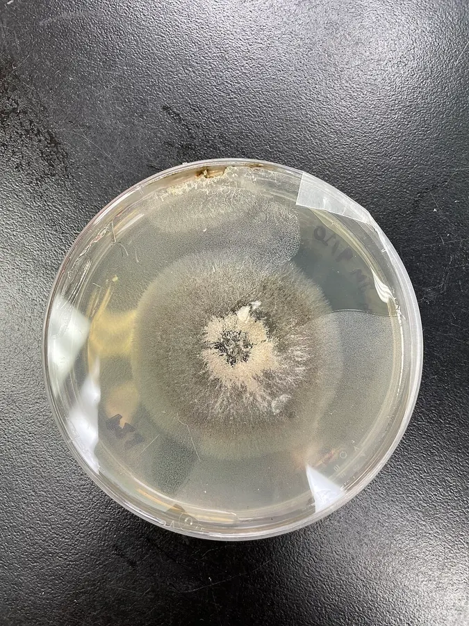
When within a host tree, A. roeperi appears as a dark stain along the walls of X. crassiusculus galleries, not immediately distinguishable from many other ambrosia fungi. Culturing A. roeperi on malt yeast extract agar (MYEA) for approximately a week in darkness at 25 °C yields large colonies with translucent, filamentous edges, immediately preceded by a dark inner ring speckled with white powder, which grows increasingly dense moving towards the middle of the colony but tapers out towards the very center. There may also be droplets of orange exudate coming from the powdery region of growth; this fluid may darken as the culture ages. The bottom of the colony similarly becomes a much deeper brown as it matures, even in refrigerated cultures. All Ambrosiella produce translucent, flask-shaped conidiogenous cells (phialides) borne on non-fertile sporodochia (minute, dense cushions of nonreproductive hyphae holding up the fertile tissue). Conidia may arise from these phialides as chains – i.e., new spores are successively pushed out, with the newest growth closest to the original conidiogenous cell – or singly, as an aleurioconidium that ruptures the membrane of the mother cell and then remains a unique, differentiated terminal unit without any subsequent neighbors produced from the phialide. A. roeperi operates only through the latter mechanism, which is distinct from what is reported in A. beaveri, A. trypodendri, and A. hartigii (suggested to use the former, chain-like method). The conidia typically retain at least one adjacent spore-producing cell when broken away from the sporodochia, distinguishing A. roeperi conidiogenous structures from the less swollen, non-detachable conidiophores of A. xylebori; the original species description suggests this feature is meant to allow these units to snap off easily while X. crassiusculus is grazing upon the gallery.

Sebagian uraian ditampilkan dalam bahasa sumber; terjemahannya belum tersedia atau belum lolos pemeriksaan.

Rujukan: [1]

Pengukuran mengikuti objek, spesimen atau kultur, dan metode yang disebutkan sumber. Baca uraian asli untuk konteks lengkap.

02 / Kehidupan

Habitat dan peran ekologis

In nature, A. roeperi is found primarily on the walls of ambrosia beetle galleries within a wide variety of host trees, where it survives by degrading compounds within wood. Traditionally, although they facilitate beetle growth and reproduction in nutritionally poor xylem tissue, ambrosia fungi are considered to be ineffective agents of wood decomposition, producing enzymes primarily dedicated to degradation of xylan, glucomannan, and callose (common components of hemicellulose) rather than cellulose, mirroring similar analyses made for various saprotrophic fungi. However, no equivalent studies have been performed on A. roeperi specifically, and it is worth noting that the article often cited in support of this conclusion only examined the fungal galleries of one beetle (Xyleborinus saxenii) associated with a different symbiont, Raffaelea sulfurea. Interestingly, metabolomic studies incorporating A. roeperi indicate that when growing on wood, this fungus possesses a lipid profile more similar to non-mutualistic Ceratocystidaceae than to other ambrosia fungi, further supporting the idea that making sweeping conclusions about fungal lifestyles based on data from apparently similar convergent systems can be misleading, as well as reinforcing how much remains to be learned about this and other ambrosial species.

Sebagian uraian ditampilkan dalam bahasa sumber; terjemahannya belum tersedia atau belum lolos pemeriksaan.

Rujukan: [1]

Habitat atau konteks temuan
Informasi belum tersedia pada sumber rilis ini.
Substrat
kayu
Peran atau konteks
Pengurai
Temukan hubungan serupa
03 / Siklus hidup

Reproduksi

When within a host tree, A. roeperi appears as a dark stain along the walls of X. crassiusculus galleries, not immediately distinguishable from many other ambrosia fungi. Culturing A. roeperi on malt yeast extract agar (MYEA) for approximately a week in darkness at 25 °C yields large colonies with translucent, filamentous edges, immediately preceded by a dark inner ring speckled with white powder, which grows increasingly dense moving towards the middle of the colony but tapers out towards the very center. There may also be droplets of orange exudate coming from the powdery region of growth; this fluid may darken as the culture ages. The bottom of the colony similarly becomes a much deeper brown as it matures, even in refrigerated cultures.

Sebagian uraian ditampilkan dalam bahasa sumber; terjemahannya belum tersedia atau belum lolos pemeriksaan.

Rujukan: [1]

04 / Batas informasi

Catatan identitas dan cakupan

  • Nama diterima mengikuti Catalogue of Life Base2026-09-11; uraian mempertahankan konsep dan nama sumber aslinya.
  • Original geographic, specimen and life-cycle scope retained; source import awaits individual editorial review.
  • Impor dan terjemahan otomatis belum diperiksa editorial per profil. Struktur reproduktif yang disebutkan tidak membuktikan seluruh siklus hidup diketahui.
  • Pengukuran pada uraian mengikuti spesimen, kultur, dan metode sumber; angka tidak diperluas ke semua populasi.

Catatan distribusi dalam narasi mengikuti sumbernya. Dokumentasi spesimen atau foto mengikuti catatan sumber. Bukti wilayah Indonesia dan penilaian konservasi tidak disimpulkan dari gambar.

05 / Dokumentasi

Melihat dengan konteks

Dokumentasi Ambrosiella roeperi dari Wikimedia Commons. The ambrosia fungus Ambrosiella roeperi growing on malt yeast extract agar at 25 degrees Celsius Identifikasi mengikuti record berkas sumber.

Jenis
Fotografi
Kredit
TwigAnnihilator
Sumber media
Buka entri sumber

Gambar menunjukkan objek menurut identifikasi sumber, bukan verifikasi independen spesimen oleh Fungi. Tidak ada skala baru yang ditambahkan.

06 / Rujukan

Sumber profil

  1. [1] Ambrosiella roeperi — Wikipedia (en)Bagian sumber: Frozen revision 1373536879
    Tanggal penghimpunan sumber: 2026-10-12
    Teks sumber: CC BY-SA4.0 · Wikipedia contributors
  2. [2] Catalogue of Life Base2026-09-11 — Ambrosiella roeperiBagian sumber: ColDP NameUsage.tsv ID CPR6; accepted species; source 2073
    Tanggal penghimpunan sumber: 2026-10-12
  3. [3] Dokumentasi visual — Ambrosiella roeperiBagian sumber: Individual media record; identification follows the source, not independent determination
    Tanggal penghimpunan sumber: 2026-10-12
    Media sumber: CC BY-SA 4.0 · TwigAnnihilator

Adaptasi teks: CC BY-SA 4.0. Wikipedia contributors — Ambrosiella roeperi — Wikipedia (en)

Uraian asli dan riwayat sumber

Riwayat atau halaman sumber

Ringkasan sumber

Ambrosiella roeperi is the fungal symbiont of the granulate ambrosia beetle, Xylosandrus crassiusculus, facilitating this insect's capacity to accumulate on and damage a diverse array of woody plants from around the world. It is one of several important nutritional partners derived from order Microascales that sustain and are transported by xylomycetophagous scolytine beetles.

Revisi sumber

Morfologi sumber

When within a host tree, A. roeperi appears as a dark stain along the walls of X. crassiusculus galleries, not immediately distinguishable from many other ambrosia fungi. Culturing A. roeperi on malt yeast extract agar (MYEA) for approximately a week in darkness at 25 °C yields large colonies with translucent, filamentous edges, immediately preceded by a dark inner ring speckled with white powder, which grows increasingly dense moving towards the middle of the colony but tapers out towards the very center. There may also be droplets of orange exudate coming from the powdery region of growth; this fluid may darken as the culture ages. The bottom of the colony similarly becomes a much deeper brown as it matures, even in refrigerated cultures. All Ambrosiella produce translucent, flask-shaped conidiogenous cells (phialides) borne on non-fertile sporodochia (minute, dense cushions of nonreproductive hyphae holding up the fertile tissue). Conidia may arise from these phialides as chains – i.e., new spores are successively pushed out, with the newest growth closest to the original conidiogenous cell – or singly, as an aleurioconidium that ruptures the membrane of the mother cell and then remains a unique, differentiated terminal unit without any subsequent neighbors produced from the phialide. A. roeperi operates only through the latter mechanism, which is distinct from what is reported in A. beaveri, A. trypodendri, and A. hartigii (suggested to use the former, chain-like method). The conidia typically retain at least one adjacent spore-producing cell when broken away from the sporodochia, distinguishing A. roeperi conidiogenous structures from the less swollen, non-detachable conidiophores of A. xylebori; the original species description suggests this feature is meant to allow these units to snap off easily while X. crassiusculus is grazing upon the gallery.

Revisi sumber

Ekologi sumber

In nature, A. roeperi is found primarily on the walls of ambrosia beetle galleries within a wide variety of host trees, where it survives by degrading compounds within wood. Traditionally, although they facilitate beetle growth and reproduction in nutritionally poor xylem tissue, ambrosia fungi are considered to be ineffective agents of wood decomposition, producing enzymes primarily dedicated to degradation of xylan, glucomannan, and callose (common components of hemicellulose) rather than cellulose, mirroring similar analyses made for various saprotrophic fungi. However, no equivalent studies have been performed on A. roeperi specifically, and it is worth noting that the article often cited in support of this conclusion only examined the fungal galleries of one beetle (Xyleborinus saxenii) associated with a different symbiont, Raffaelea sulfurea. Interestingly, metabolomic studies incorporating A. roeperi indicate that when growing on wood, this fungus possesses a lipid profile more similar to non-mutualistic Ceratocystidaceae than to other ambrosia fungi, further supporting the idea that making sweeping conclusions about fungal lifestyles based on data from apparently similar convergent systems can be misleading, as well as reinforcing how much remains to be learned about this and other ambrosial species.

Revisi sumber

Struktur reproduksi sumber

When within a host tree, A. roeperi appears as a dark stain along the walls of X. crassiusculus galleries, not immediately distinguishable from many other ambrosia fungi. Culturing A. roeperi on malt yeast extract agar (MYEA) for approximately a week in darkness at 25 °C yields large colonies with translucent, filamentous edges, immediately preceded by a dark inner ring speckled with white powder, which grows increasingly dense moving towards the middle of the colony but tapers out towards the very center. There may also be droplets of orange exudate coming from the powdery region of growth; this fluid may darken as the culture ages. The bottom of the colony similarly becomes a much deeper brown as it matures, even in refrigerated cultures.

Revisi sumber

Rujukan halaman ini

Fungi. Ambrosiella roeperi. Rilis data fungi-2026-10-12-expansion-2. https://fungi-atlas.mgadib.chatgpt.site/jamur/ambrosiella-roeperi/.

Status: impor sumber; pemeriksaan editorial per profil belum selesai. Untuk klaim biologis, gunakan rujukan pada bagian terkait dan periksa konteks sumber aslinya.