Ciri dan struktur
Spores 7.5-11 x 5-8 (9 x 6.2) µm, Q = 1.45, rather variable in size and shape, ovate to ellipsoidal, hyaline, with smooth walls and granular contents, contents becoming brownish or blue-green in KOH, inamyloid, not dextrinoid, acyanophilic. Basidia 2050 x 7-10 µm, clavate, 4-spored with conspicuous sterigmata-7 µm in length; basidioles common, like the basidia but 2.5-3 µm wide. Only a few basidia appear to mature at the same time. Cheilocystidia and pleurocystidia as usually recognised are absent but the basidia are frequently associated, both on the lamellar face and edge, with hyphoid cystidia about the same length as the basidia but narrower and with diverticulate branching towards the apex, arising at the base of the basidia or within the subhymenium (the pseudophyses of Singer & Gamundi 1963?). Trama of uniform interwoven to subparallel, hyaline, slightly thick-walled, glassy-looking hyphae, 3-6 µm diam., some with carbonaceous granules on their walls which become greenish blobs in KOH and eventually dissolve, making most of the trama and hymenium blue-green. Subhymenium of fairly closely interwoven hyphae like the trama but more closely septate and not sharply delimited from the trama. Context of similar, arrow, thickened hyphae, more branched and aboriform, but becoming thinner-walled near the pileipellis with green-staining incrustations. Pileipellis of repent hyphae giving rise to a turf of very narrow (1 -3 µm diam.) conspicuously clamped hyphae of Rameales structure, with green-staining granules, forming a network over the surface of the pileus. Stipe, when present, has similar but smaller diverticulate hyphae, some with noticeably thicker walls, arising from repent layer of slightly banded hyphae.
Habitat dan peran ekologis
Anthracophyllum archeri grows on dead wood, mostly those of angiosperms. The fungus is found in native forests throughout New Zealand,
- Habitat atau konteks temuan
- hutan
- Substrat
- kayu
- Peran atau konteks
- Belum dikategorikan pada rilis ini.
Reproduksi
Spores 7.5-11 x 5-8 (9 x 6.2) µm, Q = 1.45, rather variable in size and shape, ovate to ellipsoidal, hyaline, with smooth walls and granular contents, contents becoming brownish or blue-green in KOH, inamyloid, not dextrinoid, acyanophilic. Basidia 2050 x 7-10 µm, clavate, 4-spored with conspicuous sterigmata-7 µm in length; basidioles common, like the basidia but 2.5-3 µm wide. Only a few basidia appear to mature at the same time. Cheilocystidia and pleurocystidia as usually recognised are absent but the basidia are frequently associated, both on the lamellar face and edge, with hyphoid cystidia about the same length as the basidia but narrower and with diverticulate branching towards the apex, arising at the base of the basidia or within the subhymenium (the pseudophyses of Singer & Gamundi 1963?).
Catatan identitas dan cakupan
- Nama diterima mengikuti Catalogue of Life Base2026-09-11; uraian mempertahankan konsep dan nama sumber aslinya.
- Uraian digabungkan dari sumber spesies yang sama; lingkup geografis, bahan, dan kultur setiap sumber dipertahankan. Historical description as cited by BiotaNZ. Preserve explicit New Zealand/locality, host and specimen qualifiers. Distribution is the cited source scope, not a current global range. Original geographic, specimen and life-cycle scope retained; source import awaits individual editorial review.
- Impor dan terjemahan otomatis belum diperiksa editorial per profil. Struktur reproduktif yang disebutkan tidak membuktikan seluruh siklus hidup diketahui.
- Pengukuran pada uraian mengikuti spesimen, kultur, dan metode sumber; angka tidak diperluas ke semua populasi.
Catatan distribusi dalam narasi mengikuti sumbernya. Dokumentasi spesimen atau foto mengikuti catatan sumber. Bukti wilayah Indonesia dan penilaian konservasi tidak disimpulkan dari gambar.
Melihat dengan konteks
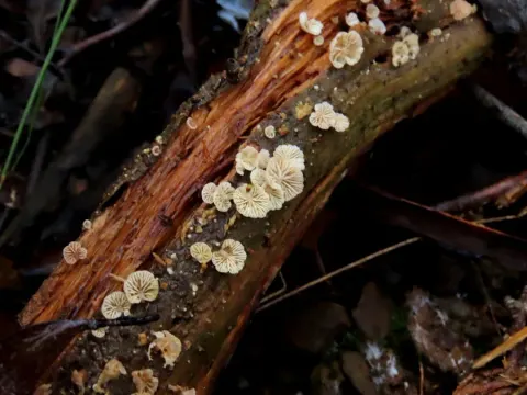
Dokumentasi Anthracophyllum archeri menurut asosiasi foto pada record takson iNaturalist. Kredit: Jerry Cooper. Identitas mengikuti sumber; bukan penentuan spesimen independen.
- Jenis
- Fotografi
- Kredit
- (c) Jerry Cooper, some rights reserved (CC BY), uploaded by Jerry Cooper
- Lisensi
- CC BY 4.0
- Sumber media
- Buka entri sumber
Dokumentasi tambahan
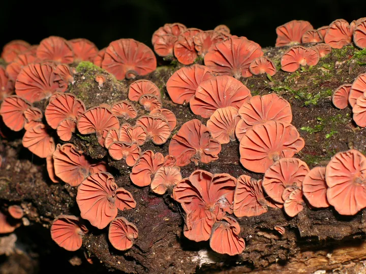
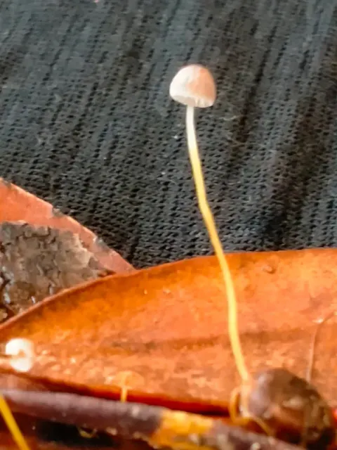
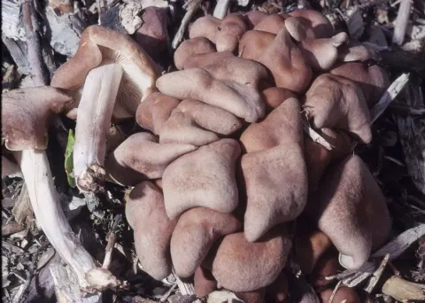
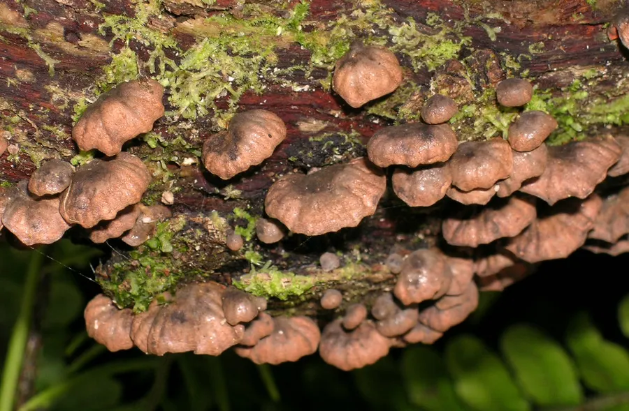
Foto yang diasosiasikan dengan Anthracophyllum archeri pada iNaturalist. Identifikasi mengikuti sumber; bukan penentuan spesimen independen.
Kredit dan penggunaan
(c) Jerry Cooper, some rights reserved (CC BY), uploaded by Jerry Cooper
Ukuran dan format gambar disesuaikan tanpa pemotongan, retus, atau penambahan skala. Lisensi sumber dipertahankan.
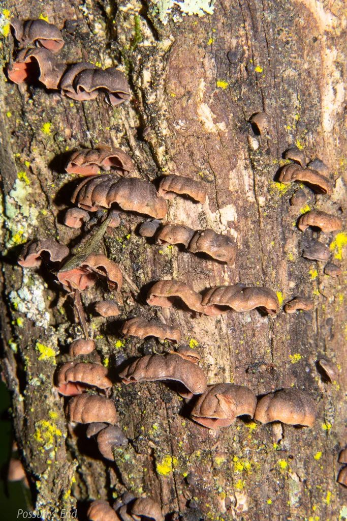
Foto yang diasosiasikan dengan Anthracophyllum archeri pada iNaturalist. Identifikasi mengikuti sumber; bukan penentuan spesimen independen.
Kredit dan penggunaan
(c) Possums' End, some rights reserved (CC BY), uploaded by Possums' End
Ukuran dan format gambar disesuaikan tanpa pemotongan, retus, atau penambahan skala. Lisensi sumber dipertahankan.
Sumber profil
- [1] Segedin, B.P. 1994: Studies in the Agaricales of New Zealand: new records and new species of the genera Cheimonophyllum, Mniopetalum, and Anthracophyllum (Tricholomataceae, Collybieae). New Zealand Journal of Botany 32: 61-72. — Anthracophyllum archeri
- [2] Anthracophyllum archeri — Wikipedia (en)
- [3] Catalogue of Life Base2026-09-11 — Anthracophyllum archeri
- [4] Dokumentasi visual — Anthracophyllum archeri
- [5] Dokumentasi visual — Anthracophyllum archeri
- [6] Dokumentasi visual — Anthracophyllum archeri
Rujukan halaman ini
Fungi. Anthracophyllum archeri. Rilis data fungi-2026-10-12-expansion-2. https://fungi-atlas.mgadib.chatgpt.site/jamur/anthracophyllum-archeri/.
Status: impor sumber; pemeriksaan editorial per profil belum selesai. Untuk klaim biologis, gunakan rujukan pada bagian terkait dan periksa konteks sumber aslinya.