Ciri dan struktur
Asexual morph: On WA, hyphae smooth, branched, septate, 1.2–3.5 µm diam. (n = 30). Conidiophores cylindrical, septate, erect, sometimes reduced to conidiogenous cells. Conidiogenous cells erect, subglobose to ampulliform, aggregated in clusters on hyphae, smooth, branched, 5–15 × 1–2.5 µm (x = 8.5 × 6 µm, n = 30). Conidia globose, sub-globose to ampulliform, lenticular, occasionally elongated to ellipsoidal, with a longitudinal germ slit, brown to dark brown, smooth to finely roughened, 6–14 × 4–7 µm (x = 10.5 × 6 µm, n = 30) µm. Sexual morph: Not observed. Colonies on PDA attaining 5 cm diam, after 4 days at 25 °C, thick, dense, surface with patches of grey aerial mycelia, margin irregular and undulate, diffuse yellow pigment, reverse yellow.
Habitat dan peran ekologis
Colonies on PDA attaining 5 cm diam, after 4 days at 25 °C, thick, dense, surface with patches of grey aerial mycelia, margin irregular and undulate, diffuse yellow pigment, reverse yellow. In the present study, two new isolates (GUCC 6.1 and GUCC 6.2) clustered together with Ap. arundinis (CBS 133509) with high-support values (ML / BI = 98/1) in the multi-locus phylogenetic tree (Fig. 1). Morphologically, GUCC 6.1 and GUCC 6.2 have similar conidiophores, conidiogenous cells, and conidia to Ap. arundinis (Li et al. 2023). Ap. arundinis was found on various plants, including Phyllostachys praecox, Castanea mollissima, and Brunfelsia brasiliensis in China (Chen et al. 2014; Liao et al. 2022; Li et al. 2023). The conidia sizes of our collection (6–14 × 4–7 µm) larger than Chen et al. (2014) (5–7 × 2–4 µm) and Liao et al. (2022) (4.5–7.4 × 3.3–4.4 µm). Comparing with the description from Li et al. (2023) (6.4–10.4 × 5.2–8.3 µm), they have similar sizes, but the conidia in this study are slenderer and more elongated. Combining phylogenetic tree and morphology, these strains were identified as Ap. arundinis.
- Habitat atau konteks temuan
- kultur yang didokumentasikan
- Substrat
- Informasi belum tersedia pada sumber rilis ini.
- Peran atau konteks
- Belum dikategorikan pada rilis ini.
Reproduksi
Asexual morph: On WA, hyphae smooth, branched, septate, 1.2–3.5 µm diam. (n = 30). Conidiophores cylindrical, septate, erect, sometimes reduced to conidiogenous cells. Conidiogenous cells erect, subglobose to ampulliform, aggregated in clusters on hyphae, smooth, branched, 5–15 × 1–2.5 µm (x = 8.5 × 6 µm, n = 30). Conidia globose, sub-globose to ampulliform, lenticular, occasionally elongated to ellipsoidal, with a longitudinal germ slit, brown to dark brown, smooth to finely roughened, 6–14 × 4–7 µm (x = 10.5 × 6 µm, n = 30) µm. Sexual morph: Not observed.
Catatan identitas dan cakupan
- Nama diterima mengikuti Catalogue of Life Base2026-09-11; uraian mempertahankan konsep dan nama sumber aslinya.
- Preserve original treatment localities, hosts and substrate scope. Materials examined describe documented specimens, not the full global range.
- Impor dan terjemahan otomatis belum diperiksa editorial per profil. Struktur reproduktif yang disebutkan tidak membuktikan seluruh siklus hidup diketahui.
- Pengukuran pada uraian mengikuti spesimen, kultur, dan metode sumber; angka tidak diperluas ke semua populasi.
Catatan distribusi dalam narasi mengikuti sumbernya. Dokumentasi spesimen atau foto mengikuti catatan sumber. Bukti wilayah Indonesia dan penilaian konservasi tidak disimpulkan dari gambar.
Melihat dengan konteks
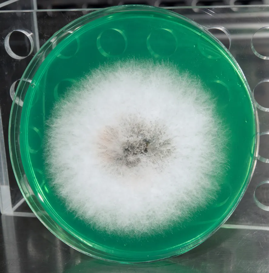
Dokumentasi Apiospora arundinis menurut asosiasi foto pada record takson iNaturalist. Kredit: Alan Rockefeller. Identitas mengikuti sumber; bukan penentuan spesimen independen.
- Jenis
- Fotografi
- Kredit
- (c) Alan Rockefeller, some rights reserved (CC BY), uploaded by Alan Rockefeller
- Lisensi
- CC BY 4.0
- Sumber media
- Buka entri sumber
Dokumentasi tambahan
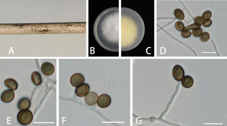
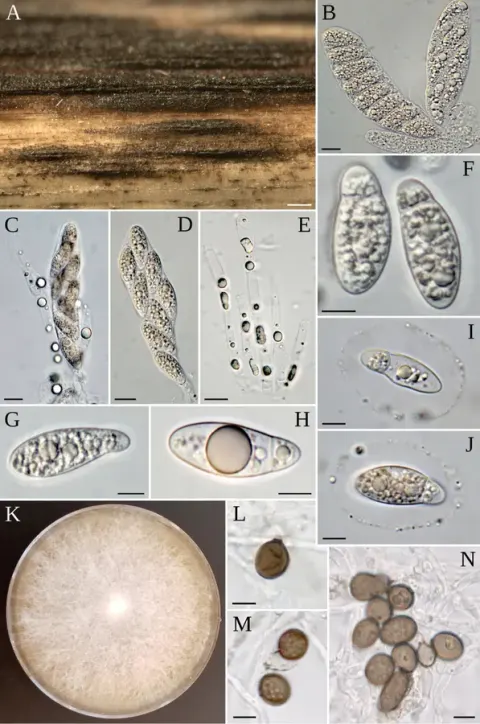
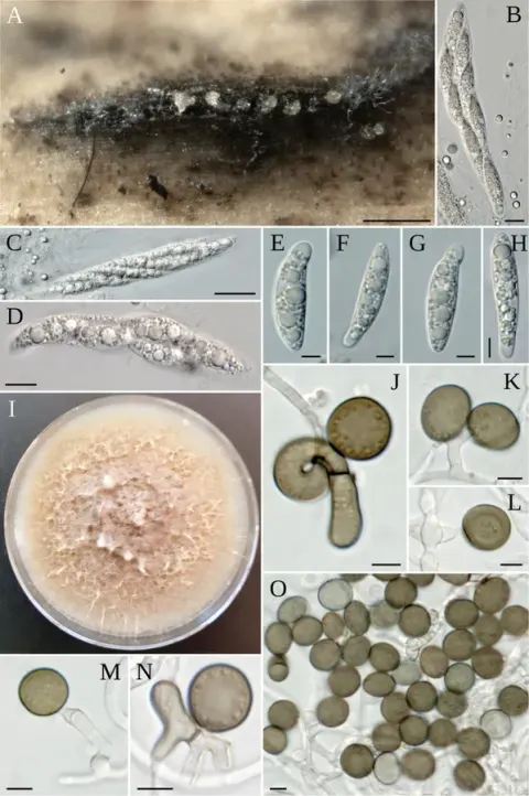
Apiospora arundinis (GUCC 6.1) A dead bamboo branch with fungi B, C above and below view of the colony on PDA D, E conidia F, G conidiophores and conidiogenous cells. Scale bars: 10 µm (D–G).
Kredit dan penggunaan
Kunmin Yu, Hong Zhang, Kexin Cheng, Yulan Jiang
Ukuran dan format gambar disesuaikan tanpa pemotongan, retus, atau penambahan skala. Lisensi sumber dipertahankan.
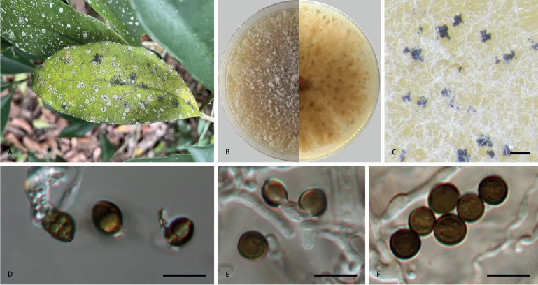
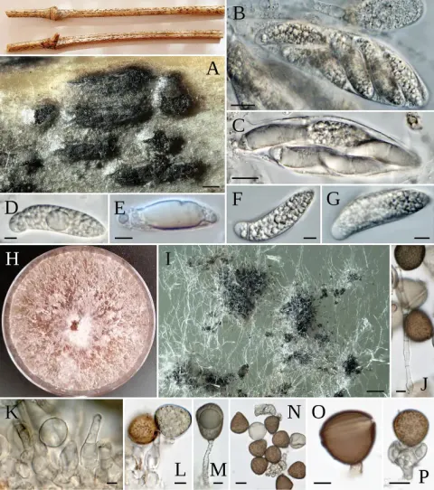
Apiospora arundinis (CFCC 58977) A leaf of host plant B colony on PDA C conidiomata formed in culture D, E conidiogenous cells giving rise to conidia F conidia. Scale bars: 1000 µm (C); 10 µm (D–F).
Kredit dan penggunaan
Shuji Li, Cheng Peng, Rong Yuan, Chengming Tian
Ukuran dan format gambar disesuaikan tanpa pemotongan, retus, atau penambahan skala. Lisensi sumber dipertahankan.
Sumber profil
- [1] Three new species of Apiospora (Amphisphaeriales, Apiosporaceae) in China — Apiospora arundinis
- [2] Catalogue of Life Base2026-09-11 — Apiospora arundinis
- [3] Dokumentasi visual — Apiospora arundinis
- [4] Dokumentasi visual — Apiospora arundinis
- [5] Dokumentasi visual — Apiospora arundinis
Rujukan halaman ini
Fungi. Apiospora arundinis. Rilis data fungi-2026-10-12-expansion-2. https://fungi-atlas.mgadib.chatgpt.site/jamur/apiospora-arundinis/.
Status: impor sumber; pemeriksaan editorial per profil belum selesai. Untuk klaim biologis, gunakan rujukan pada bagian terkait dan periksa konteks sumber aslinya.