01 / Morfologi

Ciri dan struktur

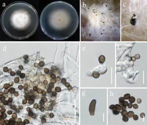
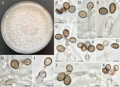
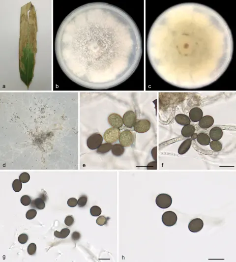
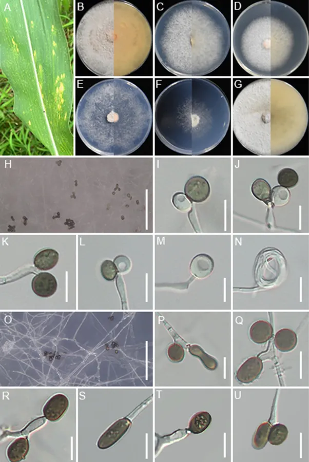
Associated with the leaf spots of maize. Lesions are yellow spots with pale brown halos on leaves, subglobose and spindle-shaped in shape, measuring 300–1000 μm in diam. Sexual morph: Not observed. Asexual morph: on WA, Hyphae 2.1–4.1 μm wide, occasionally coiled, septate, branched hyphae, hyaline. Conidiophores reduced to conidiogenous cells. Conidiogenous cells 3.9–5.1 × 1.9–3.2 μm (x̄ = 4.2 × 2.1 μm, n = 30), subglobose to ampulliform, mostly polyblastic, hyaline to pale green. Conidia 4.2–10.1 × 3.9–8.7 μm (x̄ = 7.7 × 5.9 μm, n = 30), globose to subglobose, smooth-walled, pale green when immature, brown when mature. On SNA, Conidiophores reduced to conidiogenous cells. Conidiogenous cells 3.5–8.2 × 2.1–4.2 μm (x̄ = 4.9× 2.7 μm, n = 30), subglobose to ampulliform, mostly polyblastic, pale green. Conidia 4.8–13.9 × 3.7–7.6 μm (x̄ = 8.2× 6.2 μm, n = 30), globose, subcylindrical to ovate, smooth, brown. After 7 days at 25°C, on PDA, colonies reach 88–90 mm in diam., flat, cottony, with regular margins; surface pale straw, reverse salmon. On MEA, colonies reach 70–74 mm diam., flat, with regular margins, surface, and reverse white. On CMA, colonies reaching 56–58 mm in diam., flat, with regular margins, surface, and reverse white. On SNA, colonies reach 77–79 mm in diam., flat, spreading, with scant aerial mycelia, and a regular margin, surface, and reverse that are white. On WA, colonies reach 42–45 mm diam., flat, spreading, with irregular margins, aerial mycelia scant, surface and reverse white. On OA, colonies reach 81–84 mm diam., circular, flat, cottony, with regular margin, surface white and reverse pale saffron.

Sebagian uraian ditampilkan dalam bahasa sumber; terjemahannya belum tersedia atau belum lolos pemeriksaan.

Rujukan: [1]

Pengukuran mengikuti objek, spesimen atau kultur, dan metode yang disebutkan sumber. Baca uraian asli untuk konteks lengkap.

02 / Kehidupan

Habitat dan peran ekologis

On hive-stored pollen, Hubei Province, China (Zhao et al. 2018); Aristolochia debilis, China (Chen et al. 2021); Bamboo, China (Monkai et al. 2022); Musa sp., Thailand (Samarakoon et al. 2024); grass, China (Gao et al. 2025).

Sebagian uraian ditampilkan dalam bahasa sumber; terjemahannya belum tersedia atau belum lolos pemeriksaan.

Rujukan: [1]

Habitat atau konteks temuan
Informasi belum tersedia pada sumber rilis ini.
Substrat
Informasi belum tersedia pada sumber rilis ini.
Peran atau konteks
Belum dikategorikan pada rilis ini.
Temukan hubungan serupa
03 / Siklus hidup

Reproduksi

Associated with the leaf spots of maize. Lesions are yellow spots with pale brown halos on leaves, subglobose and spindle-shaped in shape, measuring 300–1000 μm in diam. Sexual morph: Not observed. Asexual morph: on WA, Hyphae 2.1–4.1 μm wide, occasionally coiled, septate, branched hyphae, hyaline. Conidiophores reduced to conidiogenous cells. Conidiogenous cells 3.9–5.1 × 1.9–3.2 μm (x̄ = 4.2 × 2.1 μm, n = 30), subglobose to ampulliform, mostly polyblastic, hyaline to pale green. Conidia 4.2–10.1 × 3.9–8.7 μm (x̄ = 7.7 × 5.9 μm, n = 30), globose to subglobose, smooth-walled, pale green when immature, brown when mature. On SNA, Conidiophores reduced to conidiogenous cells. Conidiogenous cells 3.5–8.2 × 2.1–4.2 μm (x̄ = 4.9× 2.7 μm, n = 30), subglobose to ampulliform, mostly polyblastic, pale green.

Sebagian uraian ditampilkan dalam bahasa sumber; terjemahannya belum tersedia atau belum lolos pemeriksaan.

Rujukan: [1]

Sinonim menurut acuan taksonomi rilis: Arthrinium locutum-pollinis
04 / Batas informasi

Catatan identitas dan cakupan

  • Nama diterima mengikuti Catalogue of Life Base2026-09-11; uraian mempertahankan konsep dan nama sumber aslinya.
  • Preserve original treatment localities, hosts and substrate scope. Materials examined describe documented specimens, not the full global range.
  • Impor dan terjemahan otomatis belum diperiksa editorial per profil. Struktur reproduktif yang disebutkan tidak membuktikan seluruh siklus hidup diketahui.
  • Pengukuran pada uraian mengikuti spesimen, kultur, dan metode sumber; angka tidak diperluas ke semua populasi.

Catatan distribusi dalam narasi mengikuti sumbernya. Dokumentasi spesimen atau foto mengikuti catatan sumber. Bukti wilayah Indonesia dan penilaian konservasi tidak disimpulkan dari gambar.

05 / Dokumentasi

Melihat dengan konteks

Apiospora locuta-pollinis (GUCC 25-0071). A Appearance of the fungus on leaves of maize; B–G Upper view and reverse view of culture on PDA, MEA, CMA, SNA, WA, and OA after 7 days; H, O Close up of WA; H and SNA; O cultures with conidia; I–M Conidia with conidiogenous cells on WA; N coiled hyphae on WA; P–U Conidia with conidiogenous cells on SNA. Scale bars: 100 μm (H, O); 10 μm (I–N, P–U).

Jenis
Pelat ilmiah
Kredit
Xing-Chang Wang, Rui-Nan Zhang, Chada Norphanphoun, Si-Bo Wang, Meng-Ting Zou, Shi-Qi Guo, Jing-E Sun, Jia-Ping Wang, Nalin N. Wijayawardene, Kevin D. Hyde, Yong Wang, Feng-Quan Liu
Lisensi
CC BY 4.0
Sumber media
Buka entri sumber

Gambar menunjukkan objek menurut identifikasi sumber, bukan verifikasi independen spesimen oleh Fungi. Tidak ada skala baru yang ditambahkan.

06 / Rujukan

Sumber profil

  1. [1] Novel species and new host records of Apiospora and Nigrospora (Amphisphaeriales, Apiosporaceae) from Yunnan-Guizhou Plateau, China — Apiospora locuta-pollinisBagian sumber: PMC13135200_section_24
    Tanggal penghimpunan sumber: 2026-10-12
    Teks sumber: CC BY 4.0 · Wang XC, Zhang RN, Norphanphoun C, Wang SB, Zou MT, Guo SQ, Sun JE, Wang JP, Wijayawardene NN, Hyde KD, Wang Y, Liu FQ.
  2. [2] Catalogue of Life Base2026-09-11 — Apiospora locuta-pollinisBagian sumber: ColDP NameUsage.tsv ID 9YNCL; accepted species; source 2073
    Tanggal penghimpunan sumber: 2026-10-12
  3. [3] Dokumentasi visual — Apiospora locuta-pollinisBagian sumber: Individual media record; identification follows the source, not independent determination
    Tanggal penghimpunan sumber: 2026-10-12
    Media sumber: CC BY 4.0 · Xing-Chang Wang, Rui-Nan Zhang, Chada Norphanphoun, Si-Bo Wang, Meng-Ting Zou, Shi-Qi Guo, Jing-E Sun, Jia-Ping Wang, Nalin N. Wijayawardene, Kevin D. Hyde, Yong Wang, Feng-Quan Liu

Adaptasi teks: CC BY 4.0. Wang XC, Zhang RN, Norphanphoun C, Wang SB, Zou MT, Guo SQ, Sun JE, Wang JP, Wijayawardene NN, Hyde KD, Wang Y, Liu FQ. — Novel species and new host records of Apiospora and Nigrospora (Amphisphaeriales, Apiosporaceae) from Yunnan-Guizhou Plateau, China — Apiospora locuta-pollinis

Uraian asli dan riwayat sumber

Riwayat atau halaman sumber

Ringkasan sumber

Associated with the leaf spots of maize. Lesions are yellow spots with pale brown halos on leaves, subglobose and spindle-shaped in shape, measuring 300–1000 μm in diam. Sexual morph: Not observed. Asexual morph: on WA, Hyphae 2.1–4.1 μm wide, occasionally coiled, septate, branched hyphae, hyaline. Conidiophores reduced to conidiogenous cells. Conidiogenous cells 3.9–5.1 × 1.9–3.2 μm (x̄ = 4.2 × 2.1 μm, n = 30), subglobose to ampulliform, mostly polyblastic, hyaline to pale green. Conidia 4.2–10.1 × 3.9–8.7 μm (x̄ = 7.7 × 5.9 μm, n = 30), globose to subglobose, smooth-walled, pale green when immature, brown when mature.

Revisi sumber

Morfologi sumber

Associated with the leaf spots of maize. Lesions are yellow spots with pale brown halos on leaves, subglobose and spindle-shaped in shape, measuring 300–1000 μm in diam. Sexual morph: Not observed. Asexual morph: on WA, Hyphae 2.1–4.1 μm wide, occasionally coiled, septate, branched hyphae, hyaline. Conidiophores reduced to conidiogenous cells. Conidiogenous cells 3.9–5.1 × 1.9–3.2 μm (x̄ = 4.2 × 2.1 μm, n = 30), subglobose to ampulliform, mostly polyblastic, hyaline to pale green. Conidia 4.2–10.1 × 3.9–8.7 μm (x̄ = 7.7 × 5.9 μm, n = 30), globose to subglobose, smooth-walled, pale green when immature, brown when mature. On SNA, Conidiophores reduced to conidiogenous cells. Conidiogenous cells 3.5–8.2 × 2.1–4.2 μm (x̄ = 4.9× 2.7 μm, n = 30), subglobose to ampulliform, mostly polyblastic, pale green. Conidia 4.8–13.9 × 3.7–7.6 μm (x̄ = 8.2× 6.2 μm, n = 30), globose, subcylindrical to ovate, smooth, brown. After 7 days at 25°C, on PDA, colonies reach 88–90 mm in diam., flat, cottony, with regular margins; surface pale straw, reverse salmon. On MEA, colonies reach 70–74 mm diam., flat, with regular margins, surface, and reverse white. On CMA, colonies reaching 56–58 mm in diam., flat, with regular margins, surface, and reverse white. On SNA, colonies reach 77–79 mm in diam., flat, spreading, with scant aerial mycelia, and a regular margin, surface, and reverse that are white. On WA, colonies reach 42–45 mm diam., flat, spreading, with irregular margins, aerial mycelia scant, surface and reverse white. On OA, colonies reach 81–84 mm diam., circular, flat, cottony, with regular margin, surface white and reverse pale saffron.

Revisi sumber

Ekologi sumber

On hive-stored pollen, Hubei Province, China (Zhao et al. 2018); Aristolochia debilis, China (Chen et al. 2021); Bamboo, China (Monkai et al. 2022); Musa sp., Thailand (Samarakoon et al. 2024); grass, China (Gao et al. 2025).

Revisi sumber

Struktur reproduksi sumber

Associated with the leaf spots of maize. Lesions are yellow spots with pale brown halos on leaves, subglobose and spindle-shaped in shape, measuring 300–1000 μm in diam. Sexual morph: Not observed. Asexual morph: on WA, Hyphae 2.1–4.1 μm wide, occasionally coiled, septate, branched hyphae, hyaline. Conidiophores reduced to conidiogenous cells. Conidiogenous cells 3.9–5.1 × 1.9–3.2 μm (x̄ = 4.2 × 2.1 μm, n = 30), subglobose to ampulliform, mostly polyblastic, hyaline to pale green. Conidia 4.2–10.1 × 3.9–8.7 μm (x̄ = 7.7 × 5.9 μm, n = 30), globose to subglobose, smooth-walled, pale green when immature, brown when mature. On SNA, Conidiophores reduced to conidiogenous cells. Conidiogenous cells 3.5–8.2 × 2.1–4.2 μm (x̄ = 4.9× 2.7 μm, n = 30), subglobose to ampulliform, mostly polyblastic, pale green.

Revisi sumber

Rujukan halaman ini

Fungi. Apiospora locuta-pollinis. Rilis data fungi-2026-10-12-expansion-2. https://fungi-atlas.mgadib.chatgpt.site/jamur/apiospora-locuta-pollinis/.

Status: impor sumber; pemeriksaan editorial per profil belum selesai. Untuk klaim biologis, gunakan rujukan pada bagian terkait dan periksa konteks sumber aslinya.