Ciri dan struktur
Sexual form: Not observed. Asexual form: Fruiting bodies densely distributed on dead twigs of Platycladus orientalis, mostly immersed in the host epidermis. Conidiomata pycnidial, immersed, multilocular, solitary, 406–651 μm diam., pycnidial wall composed of dark brown textura angularis in the outer layers, gradually becoming paler toward the interior, with the innermost region thin-walled and hyaline. Ostiole central, 67–122 μm diam. Conidiophores reduced to conidiogenous cells. Conidiogenous cells cylindrical, smooth, hyaline, 6.7–14.6 × 2.1–5.9 μm (av. ± S.D. = 10.5 ± 2.5 × 3.7 ± 1.1). Paraphyses long-cylindrical, occasionally swollen at the apex, 30.7–92.5 × 1.1–7.0 μm, septate, hyaline, smooth-walled, and branched. Conidia 16.1–23.0 × 8.3–14.2 μm (av. ± S.D. = 19.8 ± 1.6 × 11.1 ± 1.3), initially hyaline, gradually turning yellowish-brown, and finally dark brown at maturity, aseptate, smooth-walled. On PDA at 25 °C under dark conditions, the colony reached approximately 60 mm in diameter after 4 days of incubation, exhibiting abundant floccose aerial mycelium. After 10 days, the colony developed pale grayish-green pigmentation, later transitioning to whitish-gray and ultimately olivaceous. Sporulation commenced after 2 weeks, forming subglobose pycnidia.
Habitat dan peran ekologis
Sexual form: Not observed. Asexual form: Fruiting bodies densely distributed on dead twigs of Platycladus orientalis, mostly immersed in the host epidermis. Conidiomata pycnidial, immersed, multilocular, solitary, 406–651 μm diam., pycnidial wall composed of dark brown textura angularis in the outer layers, gradually becoming paler toward the interior, with the innermost region thin-walled and hyaline. Ostiole central, 67–122 μm diam. Conidiophores reduced to conidiogenous cells. Conidiogenous cells cylindrical, smooth, hyaline, 6.7–14.6 × 2.1–5.9 μm (av. ± S.D. = 10.5 ± 2.5 × 3.7 ± 1.1). Paraphyses long-cylindrical, occasionally swollen at the apex, 30.7–92.5 × 1.1–7.0 μm, septate, hyaline, smooth-walled, and branched. Conidia 16.1–23.0 × 8.3–14.2 μm (av. ± S.D. = 19.8 ± 1.6 × 11.1 ± 1.3), initially hyaline, gradually turning yellowish-brown, and finally dark brown at maturity, aseptate, smooth-walled. On PDA at 25 °C under dark conditions, the colony reached approximately 60 mm in diameter after 4 days of incubation, exhibiting abundant floccose aerial mycelium. After 10 days, the colony developed pale grayish-green pigmentation, later transitioning to whitish-gray and ultimately olivaceous.
- Habitat atau konteks temuan
- kultur yang didokumentasikan
- Substrat
- Informasi belum tersedia pada sumber rilis ini.
- Peran atau konteks
- Belum dikategorikan pada rilis ini.
Reproduksi
Sexual form: Not observed. Asexual form: Fruiting bodies densely distributed on dead twigs of Platycladus orientalis, mostly immersed in the host epidermis. Conidiomata pycnidial, immersed, multilocular, solitary, 406–651 μm diam., pycnidial wall composed of dark brown textura angularis in the outer layers, gradually becoming paler toward the interior, with the innermost region thin-walled and hyaline. Ostiole central, 67–122 μm diam. Conidiophores reduced to conidiogenous cells. Conidiogenous cells cylindrical, smooth, hyaline, 6.7–14.6 × 2.1–5.9 μm (av. ± S.D. = 10.5 ± 2.5 × 3.7 ± 1.1). Paraphyses long-cylindrical, occasionally swollen at the apex, 30.7–92.5 × 1.1–7.0 μm, septate, hyaline, smooth-walled, and branched.
Catatan identitas dan cakupan
- Nama diterima mengikuti Catalogue of Life Base2026-09-11; uraian mempertahankan konsep dan nama sumber aslinya.
- Preserve original treatment localities, hosts and substrate scope. Materials examined describe documented specimens, not the full global range.
- Impor dan terjemahan otomatis belum diperiksa editorial per profil. Struktur reproduktif yang disebutkan tidak membuktikan seluruh siklus hidup diketahui.
- Pengukuran pada uraian mengikuti spesimen, kultur, dan metode sumber; angka tidak diperluas ke semua populasi.
Catatan distribusi dalam narasi mengikuti sumbernya. Dokumentasi spesimen atau foto mengikuti catatan sumber. Bukti wilayah Indonesia dan penilaian konservasi tidak disimpulkan dari gambar.
Melihat dengan konteks
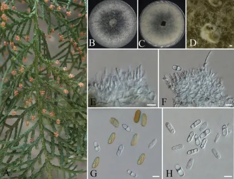
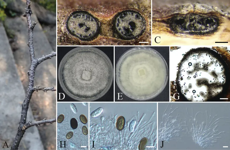
Aplosporella prunicola (CFCC 72634). A. Conidiomata on a dead twig of Platycladus orientalis; B. Transverse section of conidioma; C. Longitudinal section of conidioma; D, E. Colony morphology on PDA front and reverse views; G. Pycnidia; H. Conidia; I, J. Conidiogenous cells and paraphyses. Scale bars: 200 µm (B, C); 100 µm (G); 10 µm (H–J).
- Jenis
- Pelat ilmiah
- Kredit
- Zixian Bi, Yingying Wu, Shuji Li, Chengming Tian
- Lisensi
- CC BY 4.0
- Sumber media
- Buka entri sumber
Dokumentasi tambahan
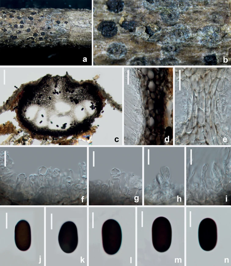
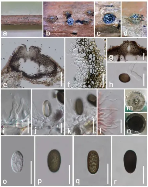
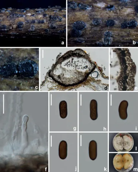
Aplosporella prunicola (HUEST 22.0091, new host record) a, b appearance of conidiomata on natural substrate c vertical section of multiloculate conidioma d, e section of peridium f–i conidiogenous cells and developing conidia j–n brown aseptate conidia. Scale bars: 100 μm (c); 20 μm (d, e); 10 μm (f–n).
Kredit dan penggunaan
Wen-Li Li, Rui-Ru Liang, Asha J. Dissanayake, Jian-Kui Liu
Ukuran dan format gambar disesuaikan tanpa pemotongan, retus, atau penambahan skala. Lisensi sumber dipertahankan.
Sumber profil
- [1] New species and records of ascomycetes on cypress in Beijing, China — Aplosporella prunicola
- [2] Catalogue of Life Base2026-09-11 — Aplosporella prunicola
- [3] Dokumentasi visual — Aplosporella prunicola
- [4] Dokumentasi visual — Aplosporella prunicola
Rujukan halaman ini
Fungi. Aplosporella prunicola. Rilis data fungi-2026-10-12-expansion-2. https://fungi-atlas.mgadib.chatgpt.site/jamur/aplosporella-prunicola/.
Status: impor sumber; pemeriksaan editorial per profil belum selesai. Untuk klaim biologis, gunakan rujukan pada bagian terkait dan periksa konteks sumber aslinya.