Ciri dan struktur
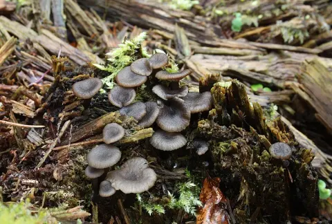
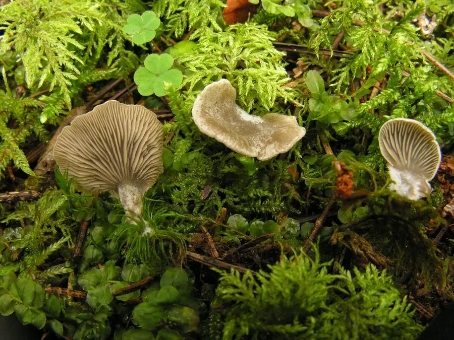
Fruit bodies (basidiocarps) of Arrhenia acerosa are small and sessile (lacking a stipe, typically fan‑shaped to kidney‑shaped (reniform) and up to 15 mm across. The thin‑fleshed cap (pileus) is smooth and medium grey‑brown, with the margin incurved when young then straightening and becoming lobed; a white, fuzzy tomentum of hyphae (thread‑like fungal cells) marks the point of attachment to the substrate. The gills (lamellae) develop from the cap margin, are of normal spacing and shape (not forked), and are slightly darker than the cap surface. Under the microscope, spores measure on average 7.4 by 3.8 μm (ranging from (5.8)6.8–8.7(9.7) by 3.4–3.9(4.4) μm), are narrowly elliptical to tear‑shaped (lacrymoid), smooth, transparent (hyaline) and bear a small apiculus (point of attachment). The spore‑bearing cells (basidia) are club‑shaped, four‑spored and measure 21–25 by 6–7 μm. No cystidia (specialised sterile cells) occur. The cap cuticle (pileipellis) is a cutis of radially orientated, cylindrical hyphae 3–10 μm wide, thin‑walled and yellow‑brown due to sparse incrustations of pigment. Clamp connections are present at the base of basidia and throughout all tissues, and pigment occurs both within cell membranes and as external incrustations.
Habitat dan peran ekologis
Arrhenia acerosa grows on decaying wood fragments or directly on bare soil among twigs and wood chips along earthy forest paths. The neotype comes from a locality in Småland, Sweden, and additional specimens have been documented in Valga County, Estonia, and Vestfold County, Norway. Its occurrence on similar substrates and in comparable habitats across these regions suggests it is a characteristic element of Northern European woodland floor communities.
- Habitat atau konteks temuan
- hutan
- Substrat
- kayu
- Peran atau konteks
- Belum dikategorikan pada rilis ini.
Reproduksi
Fruit bodies (basidiocarps) of Arrhenia acerosa are small and sessile (lacking a stipe, typically fan‑shaped to kidney‑shaped (reniform) and up to 15 mm across. The thin‑fleshed cap (pileus) is smooth and medium grey‑brown, with the margin incurved when young then straightening and becoming lobed; a white, fuzzy tomentum of hyphae (thread‑like fungal cells) marks the point of attachment to the substrate. The gills (lamellae) develop from the cap margin, are of normal spacing and shape (not forked), and are slightly darker than the cap surface. Under the microscope, spores measure on average 7.4 by 3.8 μm (ranging from (5.8)6.8–8.7(9.7) by 3.4–3.9(4.4) μm), are narrowly elliptical to tear‑shaped (lacrymoid), smooth, transparent (hyaline) and bear a small apiculus (point of attachment).
Catatan identitas dan cakupan
- Nama diterima mengikuti Catalogue of Life Base2026-09-11; uraian mempertahankan konsep dan nama sumber aslinya.
- Original geographic, specimen and life-cycle scope retained; source import awaits individual editorial review.
- Impor dan terjemahan otomatis belum diperiksa editorial per profil. Struktur reproduktif yang disebutkan tidak membuktikan seluruh siklus hidup diketahui.
- Pengukuran pada uraian mengikuti spesimen, kultur, dan metode sumber; angka tidak diperluas ke semua populasi.
Catatan distribusi dalam narasi mengikuti sumbernya. Dokumentasi spesimen atau foto mengikuti catatan sumber. Bukti wilayah Indonesia dan penilaian konservasi tidak disimpulkan dari gambar.
Melihat dengan konteks
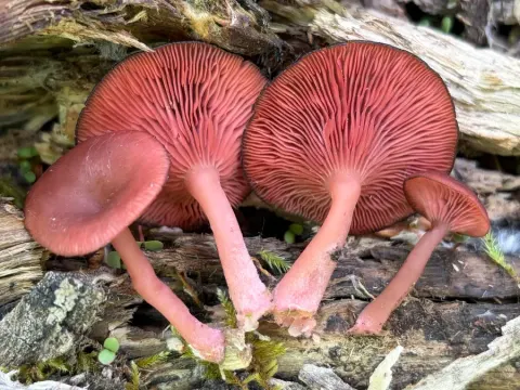
Dokumentasi Arrhenia acerosa menurut asosiasi foto pada record takson iNaturalist. Kredit: Eugene Popov. Identitas mengikuti sumber; bukan penentuan spesimen independen.
- Jenis
- Fotografi
- Kredit
- (c) Eugene Popov, some rights reserved (CC BY), uploaded by Eugene Popov
- Lisensi
- CC BY 4.0
- Sumber media
- Buka entri sumber
Sumber profil
- [1] Arrhenia acerosa — Wikipedia (en)
- [2] Catalogue of Life Base2026-09-11 — Arrhenia acerosa
- [3] Dokumentasi visual — Arrhenia acerosa
Rujukan halaman ini
Fungi. Arrhenia acerosa. Rilis data fungi-2026-10-12-expansion-2. https://fungi-atlas.mgadib.chatgpt.site/jamur/arrhenia-acerosa/.
Status: impor sumber; pemeriksaan editorial per profil belum selesai. Untuk klaim biologis, gunakan rujukan pada bagian terkait dan periksa konteks sumber aslinya.