Saprobic on mati batang udara dari Medicago sp. Conidiomata visible sebagai bulat ke oval cokelat gelap titik hitam di permukaan tuan rumah. Morph seksual tidak ditentukan. Transformasi aseksual Coelomycetous. konidiomata 100–200 μm tinggi, 80–200 μm berlian, piknidia, sepi, bercahaya atau gregaris, globosa sampai subglobosa, semi-lumut sampai terendam, kadang-kadang bermunculan, unilocular, dinding tipis, dengan papilate tunggal, ostiole yang terletak di pusat. Pycnidial walls terdiri dari lapisan 3–4, padat pada puncak dan dasar, dengan ostiol lebih tebal dari bagian lain, lapisan luar 1–2 cokelat gelap hingga hitam, lapisan dalam 1–2 hialin, dengan sel-sel dinding tipis dari tekstura angularis.
Profil ini diimpor dari uraian spesies pada sumber berlisensi dan sebagian teks diterjemahkan otomatis. Pemeriksaan editorial per profil belum selesai. Bagian yang belum diterjemahkan atau belum lolos pemeriksaan terjemahan tetap ditampilkan dalam bahasa sumber. Angka dan nama ilmiah dipertahankan.
Saprobic on dead aerial stem of Medicago sp. Conidiomata visible as round to oval dark brown to black dots on the host surface. Sexual morph Undetermined. Asexual morph Coelomycetous. Conidiomata 100–200 μm high, 80–200 μm diam., pycnidial, solitary, scattered or gregarious, globose to subglobose, semi-immersed to immersed, sometimes erumpent, unilocular, thin-walled, with a single papillate, centrally located ostiole. Pycnidial walls composed 3–4 layers, similarly dense at the apex and base, with ostiole thicker than other parts, outer 1–2 layers dark brown to black, inner 1–2 layers hyaline, with thin-walled cells of textura angularis. Conidiogenous cells 4–7 × 2–4 μm (\(\bar{x} \) = 6 × 3 µm, n = 20), hyaline, phialidic, globose. Conidia 5–10 × 1–4 μm (\(\bar{x} \) = 8 × 3 µm, n = 20), ellipsoidal to cylindrical, rounded at both ends, hyaline, straight or slightly bent at the middle, aseptate, thin- and smooth-walled, with guttulate. Culture characteristics: Colonies on PDA reaching 80 mm diam. after 7 days at 25 °C, circular colony, olivaceous to gray greenish, concentrically zonate, effuse mycelium, entire edge, reverse colony black in middle, grey olivaceous at the edge, without any diffusible pigments. Pycnidia globose to subglobose, without a distinct ostiole, glabrous, conidial matrix off whitish. Conidia sub cylindrical, aseptate.
Sebagian uraian ditampilkan dalam bahasa sumber; terjemahannya belum tersedia atau belum lolos pemeriksaan.
Pengukuran mengikuti objek, spesimen atau kultur, dan metode yang disebutkan sumber. Baca uraian asli untuk konteks lengkap.
02 / Kehidupan
Habitat dan peran ekologis
Known host and distribution: Medicago sativa (Canada Czech Republic, France, USA), Melilotus albus (Russia), Trichosanthes dioica (India) (Chen et al. 2015; Jayasiri et al. 2017; Farr and Rossman 2020).
Sebagian uraian ditampilkan dalam bahasa sumber; terjemahannya belum tersedia atau belum lolos pemeriksaan.
Saprobic on dead aerial stem of Medicago sp. Conidiomata visible as round to oval dark brown to black dots on the host surface. Sexual morph Undetermined. Asexual morph Coelomycetous. Conidiomata 100–200 μm high, 80–200 μm diam., pycnidial, solitary, scattered or gregarious, globose to subglobose, semi-immersed to immersed, sometimes erumpent, unilocular, thin-walled, with a single papillate, centrally located ostiole. Pycnidial walls composed 3–4 layers, similarly dense at the apex and base, with ostiole thicker than other parts, outer 1–2 layers dark brown to black, inner 1–2 layers hyaline, with thin-walled cells of textura angularis. Conidiogenous cells 4–7 × 2–4 μm (\(\bar{x} \) = 6 × 3 µm, n = 20), hyaline, phialidic, globose.
Sebagian uraian ditampilkan dalam bahasa sumber; terjemahannya belum tersedia atau belum lolos pemeriksaan.
Sinonim menurut acuan taksonomi rilis: Ascochyta imperfecta; Ascochyta medicaginicola var. macrospora; Phoma cuscutae; Phoma herbarum f. medicaginea; Phoma herbarum var. medicaginis; Phoma jatrophae; Phoma medicaginis; Phoma medicaginis f. microspora; Phoma medicaginis var. macrospora
04 / Batas informasi
Catatan identitas dan cakupan
Nama diterima mengikuti Catalogue of Life Base2026-09-11; uraian mempertahankan konsep dan nama sumber aslinya.
Preserve original monograph treatment localities, hosts and specimen/substrate scope. Materials examined document specimens, not the full global range.
Impor dan terjemahan otomatis belum diperiksa editorial per profil. Struktur reproduktif yang disebutkan tidak membuktikan seluruh siklus hidup diketahui.
Pengukuran pada uraian mengikuti spesimen, kultur, dan metode sumber; angka tidak diperluas ke semua populasi.
Catatan distribusi dalam narasi mengikuti sumbernya. Dokumentasi spesimen atau foto mengikuti catatan sumber. Bukti wilayah Indonesia dan penilaian konservasi tidak disimpulkan dari gambar.
05 / Dokumentasi
Melihat dengan konteks
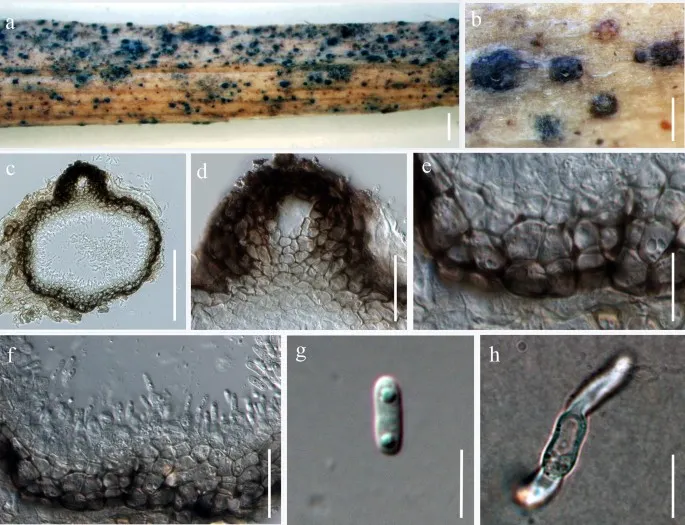
Ascochyta medicaginicola (MFLU 17- 0826). a Appearance of fruiting bodies on host substrate. b Close up view of conidiomata. c Section through conidioma. d Close up of ostiole. e Section through pycnidial wall. f Immature and mature conidia attached to conidiogenous cells. g Conidium. h Germinated conidium. Scale bars: a = 1000 µm, b = 200 µm, c = 50 µm, d – h = 10 µm
Jenis
Pelat ilmiah
Kredit
Penulis dan sumber artikel: Hyde, Kevin D.; Dong, Yang; Phookamsak, Rungtiwa; Jeewon, Rajesh; Bhat, D. Jayarama; Jones, E. B. Gareth; Liu, Ning-Guo; Abeywickrama, Pranami D.; Mapook, Ausana; Wei, Deping; Perera, Rekhani H.; Manawasinghe, Ishara S.; Pem, Dhandevi; Bundhun, Digvijayini; Karunarathna, Anuruddha; Ekanayaka, Anusha H.; Bao, Dan-Feng; Li, Junfu; Samarakoon, Milan C.; Chaiwan, Napalai; Lin, Chuan-Gen; Phutthacharoen, Kunthida; Zhang, Sheng-Nan; Senanayake, Indunil C.; Goonasekara, Ishani D.; Thambugala, Kasun M.; Phukhamsakda, Chayanard; Tennakoon, Danushka S.; Jiang, Hong-Bo; Yang, Jing; Zeng, Ming; Huanraluek, Naruemon; Liu, Jian-Kui (Jack); Wijesinghe, Subodini N.; Tian, Qing; Tibpromma, Saowaluck; Brahmanage, Rashika S.; Boonmee, Saranyaphat; Huang, Shi-Ke; Thiyagaraja, Vinodhini; Lu, Yong-Zhong; Jayawardena, Ruvishika S.; Dong, Wei; Yang, Er-Fu; Singh, Sanjay K.; Singh, Shiv Mohan; Rana, Shiwali; Lad, Sneha S.; Anand, Garima; Devadatha, Bandarupalli; Niranjan, M.; Sarma, V. Venkateswara; Liimatainen, Kare; Aguirre-Hudson, Begoña; Niskanen, Tuula; Overall, Andy; Alvarenga, Renato Lúcio Mendes; Gibertoni, Tatiana Baptista; Pfliegler, Walter P.; Horváth, Enikő; Imre, Alexandra; Alves, Amanda Lucia; da Silva Santos, Ana Carla; Tiago, Patricia Vieira; Bulgakov, Timur S.; Wanasinghe, Dhanushaka N.; Bahkali, Ali H.; Doilom, Mingkwan; Elgorban, Abdallah M.; Maharachchikumbura, Sajeewa S. N.; Rajeshkumar, Kunhiraman C.; Haelewaters, Danny; Mortimer, Peter E.; Zhao, Qi; Lumyong, Saisamorn; Xu, Jianchu; Sheng, Jun
Saprobic on dead aerial stem of Medicago sp.
Conidiomata visible as round to oval dark brown to black dots on the host surface.
Sexual morph Undetermined.
Asexual morph Coelomycetous.
Conidiomata 100–200 μm high, 80–200 μm diam., pycnidial, solitary, scattered or gregarious, globose to subglobose, semi-immersed to immersed, sometimes erumpent, unilocular, thin-walled, with a single papillate, centrally located ostiole.
Pycnidial walls composed 3–4 layers, similarly dense at the apex and base, with ostiole thicker than other parts, outer 1–2 layers dark brown to black, inner 1–2 layers hyaline, with thin-walled cells of textura angularis.
Saprobic on dead aerial stem of Medicago sp. Conidiomata visible as round to oval dark brown to black dots on the host surface. Sexual morph Undetermined. Asexual morph Coelomycetous. Conidiomata 100–200 μm high, 80–200 μm diam., pycnidial, solitary, scattered or gregarious, globose to subglobose, semi-immersed to immersed, sometimes erumpent, unilocular, thin-walled, with a single papillate, centrally located ostiole. Pycnidial walls composed 3–4 layers, similarly dense at the apex and base, with ostiole thicker than other parts, outer 1–2 layers dark brown to black, inner 1–2 layers hyaline, with thin-walled cells of textura angularis. Conidiogenous cells 4–7 × 2–4 μm (\(\bar{x} \) = 6 × 3 µm, n = 20), hyaline, phialidic, globose. Conidia 5–10 × 1–4 μm (\(\bar{x} \) = 8 × 3 µm, n = 20), ellipsoidal to cylindrical, rounded at both ends, hyaline, straight or slightly bent at the middle, aseptate, thin- and smooth-walled, with guttulate.
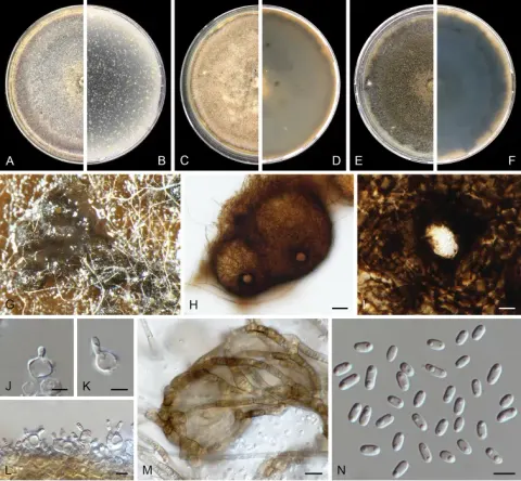
Culture characteristics: Colonies on PDA reaching 80 mm diam. after 7 days at 25 °C, circular colony, olivaceous to gray greenish, concentrically zonate, effuse mycelium, entire edge, reverse colony black in middle, grey olivaceous at the edge, without any diffusible pigments. Pycnidia globose to subglobose, without a distinct ostiole, glabrous, conidial matrix off whitish. Conidia sub cylindrical, aseptate.
Known host and distribution: Medicago sativa (Canada Czech Republic, France, USA), Melilotus albus (Russia), Trichosanthes dioica (India) (Chen et al. 2015; Jayasiri et al. 2017; Farr and Rossman 2020).
Saprobic on dead aerial stem of Medicago sp.
Conidiomata visible as round to oval dark brown to black dots on the host surface.
Sexual morph Undetermined.
Asexual morph Coelomycetous.
Conidiomata 100–200 μm high, 80–200 μm diam., pycnidial, solitary, scattered or gregarious, globose to subglobose, semi-immersed to immersed, sometimes erumpent, unilocular, thin-walled, with a single papillate, centrally located ostiole.
Pycnidial walls composed 3–4 layers, similarly dense at the apex and base, with ostiole thicker than other parts, outer 1–2 layers dark brown to black, inner 1–2 layers hyaline, with thin-walled cells of textura angularis.
Conidiogenous cells 4–7 × 2–4 μm (\(\bar{x} \) = 6 × 3 µm, n = 20), hyaline, phialidic, globose.
Status: impor sumber; pemeriksaan editorial per profil belum selesai. Untuk klaim biologis, gunakan rujukan pada bagian terkait dan periksa konteks sumber aslinya.
Koloni setelah 7 d di 25 °C, pada OA mencapai 50–55 mm diam, flukos mycelium udara, hitam zaitun hingga zaitun, buff ke peripheria, produksi banyak pycnidia, margin tidak teratur; berwarna kebalik dengan permukaan. Pada MEA mencapai 40–45…