Saprobic on daun mati dari Zea mays. Morph seksual tidak ditentukan. Transformasi aseksual Coelomycetous. konidiomata 80–110 μm tinggi, 110–140 μm diam. (\(\bar{x} \) = 126.2 × 99 μm, n = 10), pycnidial, cokelat gelap hingga hitam, tenggelam, sedikit diangkat, solitary hingga gregarious, uni-loculate, globose hingga subglobosa, glabrous, ostiolate, dengan papilla kecil. Conidiomata walls 8–20 µm lebar, dinding sedikit tebal, tebalnya sama, terdiri dari beberapa lapisan sel coklat, sel-sel pseudoparenkimat dari tekstura angularis, lebih pucat ke lapisan dalam.
Profil ini diimpor dari uraian spesies pada sumber berlisensi dan sebagian teks diterjemahkan otomatis. Pemeriksaan editorial per profil belum selesai. Bagian yang belum diterjemahkan atau belum lolos pemeriksaan terjemahan tetap ditampilkan dalam bahasa sumber. Angka dan nama ilmiah dipertahankan.
Saprobic on dead leaves of Zea mays. Sexual morph Undetermined. Asexual morph Coelomycetous. Conidiomata 80–110 μm high, 110–140 μm diam. (\(\bar{x} \) = 126.2 × 99 μm, n = 10), pycnidial, dark brown to black, immersed, slightly raised, solitary to gregarious, uni-loculate, globose to subglobose, glabrous, ostiolate, with a minute papilla. Conidiomata walls 8–20 µm wide, slightly thick-walled, of equal thickness, comprising several cell layers of brown, pseudoparenchymatous cells of textura angularis, paler towards the inner layers. Conidiophores arising from the inner cavity, reduced to conidiogenous cells. Conidiogenous cells (3.6–)4–7.5 × 2–5 μm (\(\bar{x} \) = 5.4 × 2.8 μm, n = 30), holoblastic, phialidic, rarely with 1–2 percurrent proliferations, discrete, hyaline, ampulliform to subcylindrical, or obclavate, aseptate, smooth-walled. Conidia (17.5–)20–25 × 3–4 μm (\(\bar{x} \) = 22.1 × 3.9 μm, n = 30), cylindrical to subcylindrical, straight to slightly curved, 4-septate, not constricted at the septa, with longest cell at the second from base, bearing appendages; basal cell 2.5–4 μm long (\(\bar{x} \) = 3.2 µm), obconic, truncate at base, hyaline, thin and smooth-walled, second cell from the base 6.5–8 μm long (\(\bar{x} \) = 7.4 µm), pale yellowish, third cell 4–5.5 μm long (\(\bar{x} \) = 4.8 μm), pale yellowish, fourth cell 4–5.5(–6) μm long (\(\bar{x} \) = 5 μm), pale yellowish, apical cell 2–3(–3.7) μm long (\(\bar{x} \) = 2.9 μm), conical, hyaline and smooth-walled, forming three-branched tubular, flexuous, 10–20 µm long apical appendages; basal appendages 5–6 µm long, single, absent at immature state, tubular, unbranched, centric.
Sebagian uraian ditampilkan dalam bahasa sumber; terjemahannya belum tersedia atau belum lolos pemeriksaan.
Pengukuran mengikuti objek, spesimen atau kultur, dan metode yang disebutkan sumber. Baca uraian asli untuk konteks lengkap.
02 / Kehidupan
Habitat dan peran ekologis
Saprobic on dead leaves of Zea mays. Sexual morph Undetermined. Asexual morph Coelomycetous. Conidiomata 80–110 μm high, 110–140 μm diam. (\(\bar{x} \) = 126.2 × 99 μm, n = 10), pycnidial, dark brown to black, immersed, slightly raised, solitary to gregarious, uni-loculate, globose to subglobose, glabrous, ostiolate, with a minute papilla. Conidiomata walls 8–20 µm wide, slightly thick-walled, of equal thickness, comprising several cell layers of brown, pseudoparenchymatous cells of textura angularis, paler towards the inner layers. Conidiophores arising from the inner cavity, reduced to conidiogenous cells. Conidiogenous cells (3.6–)4–7.5 × 2–5 μm (\(\bar{x} \) = 5.4 × 2.8 μm, n = 30), holoblastic, phialidic, rarely with 1–2 percurrent proliferations, discrete, hyaline, ampulliform to subcylindrical, or obclavate, aseptate, smooth-walled. Conidia (17.5–)20–25 × 3–4 μm (\(\bar{x} \) = 22.1 × 3.9 μm, n = 30), cylindrical to subcylindrical, straight to slightly curved, 4-septate, not constricted at the septa, with longest cell at the second from base, bearing appendages; basal cell 2.5–4 μm long (\(\bar{x} \) = 3.2 µm), obconic, truncate at base, hyaline, thin and smooth-walled, second cell from the base 6.5–8 μm long (\(\bar{x} \) = 7.4 µm), pale yellowish, third cell 4–5.5 μm long (\(\bar{x} \) = 4.8 μm), pale yellowish, fourth cell 4–5.5(–6) μm long (\(\bar{x} \) = 5 μm), pale yellowish, apical cell 2–3(–3.7) μm long (\(\bar{x} \) = 2.9 μm), conical, hyaline and smooth-walled, forming three-branched tubular, flexuous, 10–20 µm long apical appendages; basal appendages 5–6 µm long, single, absent at immature state, tubular, unbranched, centric.
Sebagian uraian ditampilkan dalam bahasa sumber; terjemahannya belum tersedia atau belum lolos pemeriksaan.
Saprobic on dead leaves of Zea mays. Sexual morph Undetermined. Asexual morph Coelomycetous. Conidiomata 80–110 μm high, 110–140 μm diam. (\(\bar{x} \) = 126.2 × 99 μm, n = 10), pycnidial, dark brown to black, immersed, slightly raised, solitary to gregarious, uni-loculate, globose to subglobose, glabrous, ostiolate, with a minute papilla. Conidiomata walls 8–20 µm wide, slightly thick-walled, of equal thickness, comprising several cell layers of brown, pseudoparenchymatous cells of textura angularis, paler towards the inner layers. Conidiophores arising from the inner cavity, reduced to conidiogenous cells. Conidiogenous cells (3.6–)4–7.5 × 2–5 μm (\(\bar{x} \) = 5.4 × 2.8 μm, n = 30), holoblastic, phialidic, rarely with 1–2 percurrent proliferations, discrete, hyaline, ampulliform to subcylindrical, or obclavate, aseptate, smooth-walled.
Sebagian uraian ditampilkan dalam bahasa sumber; terjemahannya belum tersedia atau belum lolos pemeriksaan.
Nama diterima mengikuti Catalogue of Life Base2026-09-11; uraian mempertahankan konsep dan nama sumber aslinya.
Preserve original monograph treatment localities, hosts and specimen/substrate scope. Materials examined document specimens, not the full global range.
Impor dan terjemahan otomatis belum diperiksa editorial per profil. Struktur reproduktif yang disebutkan tidak membuktikan seluruh siklus hidup diketahui.
Pengukuran pada uraian mengikuti spesimen, kultur, dan metode sumber; angka tidak diperluas ke semua populasi.
Catatan distribusi dalam narasi mengikuti sumbernya. Dokumentasi spesimen atau foto mengikuti catatan sumber. Bukti wilayah Indonesia dan penilaian konservasi tidak disimpulkan dari gambar.
05 / Dokumentasi
Melihat dengan konteks
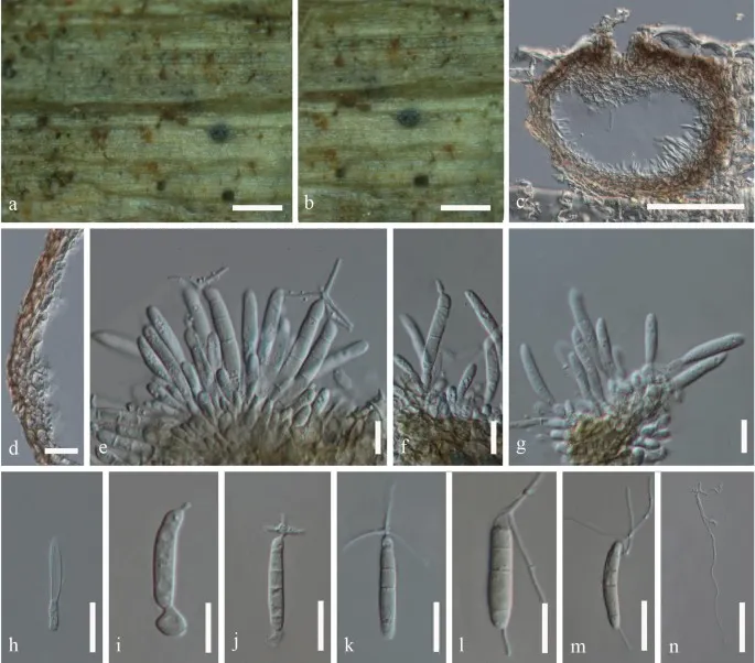
Bartalinia kunmingensis (KUN-HKAS 102242, holotype). a, b Conidiomata on host substrate. c Vertical section of conidioma. d Conidioma wall. e–i Conidiogenous cells with conidia. j–m Conidia. n Germinating conidium. Scale bars a, b = 200 μm, c = 50 μm, d–m = 10 μm, n = 20 μm
Jenis
Pelat ilmiah
Kredit
Penulis dan sumber artikel: Phookamsak, Rungtiwa; Hyde, Kevin D.; Jeewon, Rajesh; Bhat, D. Jayarama; Jones, E. B. Gareth; Maharachchikumbura, Sajeewa S. N.; Raspé, Olivier; Karunarathna, Samantha C.; Wanasinghe, Dhanushka N.; Hongsanan, Sinang; Doilom, Mingkwan; Tennakoon, Danushka S.; Machado, Alexandre R.; Firmino, André L.; Ghosh, Aniket; Karunarathna, Anuruddha; Mešić, Armin; Dutta, Arun Kumar; Thongbai, Benjarong; Devadatha, Bandarupalli; Norphanphoun, Chada; Senwanna, Chanokned; Wei, Deping; Pem, Dhandevi; Ackah, Frank Kwekucher; Wang, Gen-Nuo; Jiang, Hong-Bo; Madrid, Hugo; Lee, Hyang Burm; Goonasekara, Ishani D.; Manawasinghe, Ishara S.; Kušan, Ivana; Cano, Josep; Gené, Josepa; Li, Junfu; Das, Kanad; Acharya, Krishnendu; Raj, K. N. Anil; Latha, K. P. Deepna; Chethana, K. W. Thilini; He, Mao-Qiang; Dueñas, Margarita; Jadan, Margita; Martín, María P.; Samarakoon, Milan C.; Dayarathne, Monika C.; Raza, Mubashar; Park, Myung Soo; Telleria, M. Teresa; Chaiwan, Napalai; Matočec, Neven; de Silva, Nimali I.; Pereira, Olinto L.; Singh, Paras Nath; Manimohan, Patinjareveettil; Uniyal, Priyanka; Shang, Qiu-Ju; Bhatt, Rajendra P.; Perera, Rekhani H.; Alvarenga, Renato Lúcio Mendes; Nogal-Prata, Sandra; Singh, Sanjay K.; Vadthanarat, Santhiti; Oh, Seung-Yoon; Huang, Shi-Ke; Rana, Shiwali; Konta, Sirinapa; Paloi, Soumitra; Jayasiri, Subashini C.; Jeon, Sun Jeong; Mehmood, Tahir; Gibertoni, Tatiana Baptista; Nguyen, Thuong T. T.; Singh, Upendra; Thiyagaraja, Vinodhini; Sarma, V. Venkateswara; Dong, Wei; Yu, Xian-Dong; Lu, Yong-Zhong; Lim, Young Woon; Chen, Yun; Tkalčec, Zdenko; Zhang, Zhi-Feng; Luo, Zong-Long; Daranagama, Dinushani A.; Thambugala, Kasun M.; Tibpromma, Saowaluck; Camporesi, Erio; Bulgakov, Timur S.; Dissanayake, Asha J.; Senanayake, Indunil C.; Dai, Dong Qin; Tang, Li-Zhou; Khan, Sehroon; Zhang, Huang; Promputtha, Itthayakorn; Cai, Lei; Chomnunti, Putarak; Zhao, Rui-Lin; Lumyong, Saisamorn; Boonmee, Saranyaphat; Wen, Ting-Chi; Mortimer, Peter E.; Xu, Jianchu
Saprobic on dead leaves of Zea mays.
Sexual morph Undetermined.
Asexual morph Coelomycetous.
Conidiomata 80–110 μm high, 110–140 μm diam. (\(\bar{x} \) = 126.2 × 99 μm, n = 10), pycnidial, dark brown to black, immersed, slightly raised, solitary to gregarious, uni-loculate, globose to subglobose, glabrous, ostiolate, with a minute papilla.
Conidiomata walls 8–20 µm wide, slightly thick-walled, of equal thickness, comprising several cell layers of brown, pseudoparenchymatous cells of textura angularis, paler towards the inner layers.
Saprobic on dead leaves of Zea mays.
Sexual morph Undetermined.
Asexual morph Coelomycetous.
Conidiomata 80–110 μm high, 110–140 μm diam. (\(\bar{x} \) = 126.2 × 99 μm, n = 10), pycnidial, dark brown to black, immersed, slightly raised, solitary to gregarious, uni-loculate, globose to subglobose, glabrous, ostiolate, with a minute papilla.
Conidiomata walls 8–20 µm wide, slightly thick-walled, of equal thickness, comprising several cell layers of brown, pseudoparenchymatous cells of textura angularis, paler towards the inner layers.
Conidiophores arising from the inner cavity, reduced to conidiogenous cells.
Conidiogenous cells (3.6–)4–7.5 × 2–5 μm (\(\bar{x} \) = 5.4 × 2.8 μm, n = 30), holoblastic, phialidic, rarely with 1–2 percurrent proliferations, discrete, hyaline, ampulliform to subcylindrical, or obclavate, aseptate, smooth-walled.
Conidia (17.5–)20–25 × 3–4 μm (\(\bar{x} \) = 22.1 × 3.9 μm, n = 30), cylindrical to subcylindrical, straight to slightly curved, 4-septate, not constricted at the septa, with longest cell at the second from base, bearing appendages; basal cell 2.5–4 μm long (\(\bar{x} \) = 3.2 µm), obconic, truncate at base, hyaline, thin and smooth-walled, second cell from the base 6.5–8 μm long (\(\bar{x} \) = 7.4 µm), pale yellowish, third cell 4–5.5 μm long (\(\bar{x} \) = 4.8 μm), pale yellowish, fourth cell 4–5.5(–6) μm long (\(\bar{x} \) = 5 μm), pale yellowish, apical cell 2–3(–3.7) μm long (\(\bar{x} \) = 2.9 μm), conical, hyaline and smooth-walled, forming three-branched tubular, flexuous, 10–20 µm long apical appendages; basal appendages 5–6 µm long, single, absent at immature state, tubular, unbranched, centric.
Saprobic on dead leaves of Zea mays.
Sexual morph Undetermined.
Asexual morph Coelomycetous.
Conidiomata 80–110 μm high, 110–140 μm diam. (\(\bar{x} \) = 126.2 × 99 μm, n = 10), pycnidial, dark brown to black, immersed, slightly raised, solitary to gregarious, uni-loculate, globose to subglobose, glabrous, ostiolate, with a minute papilla.
Conidiomata walls 8–20 µm wide, slightly thick-walled, of equal thickness, comprising several cell layers of brown, pseudoparenchymatous cells of textura angularis, paler towards the inner layers.
Conidiophores arising from the inner cavity, reduced to conidiogenous cells.
Conidiogenous cells (3.6–)4–7.5 × 2–5 μm (\(\bar{x} \) = 5.4 × 2.8 μm, n = 30), holoblastic, phialidic, rarely with 1–2 percurrent proliferations, discrete, hyaline, ampulliform to subcylindrical, or obclavate, aseptate, smooth-walled.
Conidia (17.5–)20–25 × 3–4 μm (\(\bar{x} \) = 22.1 × 3.9 μm, n = 30), cylindrical to subcylindrical, straight to slightly curved, 4-septate, not constricted at the septa, with longest cell at the second from base, bearing appendages; basal cell 2.5–4 μm long (\(\bar{x} \) = 3.2 µm), obconic, truncate at base, hyaline, thin and smooth-walled, second cell from the base 6.5–8 μm long (\(\bar{x} \) = 7.4 µm), pale yellowish, third cell 4–5.5 μm long (\(\bar{x} \) = 4.8 μm), pale yellowish, fourth cell 4–5.5(–6) μm long (\(\bar{x} \) = 5 μm), pale yellowish, apical cell 2–3(–3.7) μm long (\(\bar{x} \) = 2.9 μm), conical, hyaline and smooth-walled, forming three-branched tubular, flexuous, 10–20 µm long apical appendages; basal appendages 5–6 µm long, single, absent at immature state, tubular, unbranched, centric.
Saprobic on dead leaves of Zea mays.
Sexual morph Undetermined.
Asexual morph Coelomycetous.
Conidiomata 80–110 μm high, 110–140 μm diam. (\(\bar{x} \) = 126.2 × 99 μm, n = 10), pycnidial, dark brown to black, immersed, slightly raised, solitary to gregarious, uni-loculate, globose to subglobose, glabrous, ostiolate, with a minute papilla.
Conidiomata walls 8–20 µm wide, slightly thick-walled, of equal thickness, comprising several cell layers of brown, pseudoparenchymatous cells of textura angularis, paler towards the inner layers.
Conidiophores arising from the inner cavity, reduced to conidiogenous cells.
Conidiogenous cells (3.6–)4–7.5 × 2–5 μm (\(\bar{x} \) = 5.4 × 2.8 μm, n = 30), holoblastic, phialidic, rarely with 1–2 percurrent proliferations, discrete, hyaline, ampulliform to subcylindrical, or obclavate, aseptate, smooth-walled.
Status: impor sumber; pemeriksaan editorial per profil belum selesai. Untuk klaim biologis, gunakan rujukan pada bagian terkait dan periksa konteks sumber aslinya.
Beauveria bassiana adalah jamur yang tumbuh secara alami di tanah di seluruh dunia dan bertindak sebagai parasit pada berbagai spesies arthropoda, menyebabkan penyakit muscardin putih; dengan demikian, ia termasuk dalam kelompok jamur…
Deskripsi morfologis dari penelitian ini didasarkan pada sampel, CBS 250.34. Morph seksual: tidak ditentukan. Transformasi aseksual: Koloni pada PDA mencapai diameter 36–38 mm setelah sebulan di 25 °C, putih hingga putih krim, tekstur…
Tubuh buah Cordyceps locustiphila membentuk kelompok seperti tongkat melalui celah dan sendi di kerang chitinous dari ulat tuan rumah mereka. stipe dari klub adalah kuning abu-abu daging dengan panjang 1–4 mm panjang dan diameter 1–2 mm. S…