Saprobic on cabang mati dari Coronilla emerus. Morph seksual Ascomata 370–500 × 340–430 μm (\(\bar{x} \) = 411 × 379 μm, n = 5), dangkal hingga semi-lumut, globosa hingga subglobosa, sepi, berkeliaran, mencolok di permukaan, uniloculate, hitam, ostioles, tanpa papilla. Peridium 35–80 μm lebar, berbatu tebal, terdiri dari sel-sel subyalin sampai kuning ⁇ coklat dari tekstura angularis. Hamathecium 1.5–3 μm lebar, terdiri dari banyak, filamen, bercabang, bersepta, pseudoparaphyses. askus 100–190 × 10–15 μm (\(\bar{x} \) = 158 × 13 μm, n = 20), 4(6) –8-spored, sebagian besar 8-spored, bitunikat, fissitunikat, silinder, pedicellate, dengan ruang mata kecil.
Profil ini diimpor dari uraian spesies pada sumber berlisensi dan sebagian teks diterjemahkan otomatis. Pemeriksaan editorial per profil belum selesai. Bagian yang belum diterjemahkan atau belum lolos pemeriksaan terjemahan tetap ditampilkan dalam bahasa sumber. Angka dan nama ilmiah dipertahankan.
Saprobic on dead branch of Coronilla emerus. Sexual morph Ascomata 370–500 × 340–430 μm (\(\bar{x} \) = 411 × 379 μm, n = 5), superficial to semi-immersed, globose to subglobose, solitary, scattered, conspicuous at the surface, uniloculate, black, ostioles, without papilla. Peridium 35–80 μm wide, thick-walled, comprising subhyaline to yellow–brown cells of textura angularis. Hamathecium 1.5–3 μm wide, comprising numerous, filamentous, branched, septate, pseudoparaphyses. Asci 100–190 × 10–15 μm (\(\bar{x} \) = 158 × 13 μm, n = 20), 4(6)–8-spored, mostly 8-spored, bitunicate, fissitunicate, cylindrical, pedicellate, with minute ocular chamber. Ascospores 19–25 × 8–13 μm (\(\bar{x} \) = 21 × 10 μm, n = 20), 1-seriate, sometimes overlapping, muriform, ellipsoidal to subfusiform, slightly curved, upper part wider than the lower part, 3–5 transversely septate, with 1–3 vertical septa, constricted at the central septum, slightly constricted at the septa, initially subhyaline, becoming yellow–brown to brown at maturity, with narrowly rounded ends, with guttules, thick and smooth-walled, lacking a mucilaginous sheath. Asexual morph Undetermined. Culture characteristics: Colonies on MEA at room temperature reaching 9 cm in 6–8 weeks, circular with curved edges, white mycelium raised from the medium surface, not sporulating in culture within two months.
Sebagian uraian ditampilkan dalam bahasa sumber; terjemahannya belum tersedia atau belum lolos pemeriksaan.
Pengukuran mengikuti objek, spesimen atau kultur, dan metode yang disebutkan sumber. Baca uraian asli untuk konteks lengkap.
02 / Kehidupan
Habitat dan peran ekologis
Saprobic on dead branch of Coronilla emerus. Sexual morph Ascomata 370–500 × 340–430 μm (\(\bar{x} \) = 411 × 379 μm, n = 5), superficial to semi-immersed, globose to subglobose, solitary, scattered, conspicuous at the surface, uniloculate, black, ostioles, without papilla. Peridium 35–80 μm wide, thick-walled, comprising subhyaline to yellow–brown cells of textura angularis. Hamathecium 1.5–3 μm wide, comprising numerous, filamentous, branched, septate, pseudoparaphyses. Asci 100–190 × 10–15 μm (\(\bar{x} \) = 158 × 13 μm, n = 20), 4(6)–8-spored, mostly 8-spored, bitunicate, fissitunicate, cylindrical, pedicellate, with minute ocular chamber. Ascospores 19–25 × 8–13 μm (\(\bar{x} \) = 21 × 10 μm, n = 20), 1-seriate, sometimes overlapping, muriform, ellipsoidal to subfusiform, slightly curved, upper part wider than the lower part, 3–5 transversely septate, with 1–3 vertical septa, constricted at the central septum, slightly constricted at the septa, initially subhyaline, becoming yellow–brown to brown at maturity, with narrowly rounded ends, with guttules, thick and smooth-walled, lacking a mucilaginous sheath. Asexual morph Undetermined. Material examined: ITALY, Province of Forlì-Cesena [FC], Monte Colombo-Predappio, on dead aerial branch of Coronilla emerus (L.) Lassen (Fabaceae), 25 November 2018, E.
Sebagian uraian ditampilkan dalam bahasa sumber; terjemahannya belum tersedia atau belum lolos pemeriksaan.
Saprobic on dead branch of Coronilla emerus. Sexual morph Ascomata 370–500 × 340–430 μm (\(\bar{x} \) = 411 × 379 μm, n = 5), superficial to semi-immersed, globose to subglobose, solitary, scattered, conspicuous at the surface, uniloculate, black, ostioles, without papilla. Peridium 35–80 μm wide, thick-walled, comprising subhyaline to yellow–brown cells of textura angularis. Hamathecium 1.5–3 μm wide, comprising numerous, filamentous, branched, septate, pseudoparaphyses. Asci 100–190 × 10–15 μm (\(\bar{x} \) = 158 × 13 μm, n = 20), 4(6)–8-spored, mostly 8-spored, bitunicate, fissitunicate, cylindrical, pedicellate, with minute ocular chamber. Ascospores 19–25 × 8–13 μm (\(\bar{x} \) = 21 × 10 μm, n = 20), 1-seriate, sometimes overlapping, muriform, ellipsoidal to subfusiform, slightly curved, upper part wider than the lower part, 3–5 transversely septate, with 1–3 vertical septa, constricted at the central septum, slightly constricted at the septa, initially subhyaline, becoming yellow–brown to brown at maturity, with narrowly rounded ends, with guttules, thick and smooth-walled, lacking a mucilaginous sheath.
Sebagian uraian ditampilkan dalam bahasa sumber; terjemahannya belum tersedia atau belum lolos pemeriksaan.
Nama diterima mengikuti Catalogue of Life Base2026-09-11; uraian mempertahankan konsep dan nama sumber aslinya.
Preserve original monograph treatment localities, hosts and specimen/substrate scope. Materials examined document specimens, not the full global range.
Impor dan terjemahan otomatis belum diperiksa editorial per profil. Struktur reproduktif yang disebutkan tidak membuktikan seluruh siklus hidup diketahui.
Pengukuran pada uraian mengikuti spesimen, kultur, dan metode sumber; angka tidak diperluas ke semua populasi.
Catatan distribusi dalam narasi mengikuti sumbernya. Dokumentasi spesimen atau foto mengikuti catatan sumber. Bukti wilayah Indonesia dan penilaian konservasi tidak disimpulkan dari gambar.
05 / Dokumentasi
Melihat dengan konteks
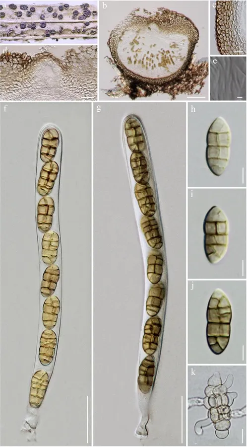
Camarosporidiella camporesii (MFLU 19-0296, holotype). a Appearance of ascomata on host substrate. b Section of ascoma. c Peridium. d Close up of ostiole. e Pseudoparaphyses. f, g Asci. h – j Ascospores. k Germinating ascospore. Scale bars: b = 100 μm, c, d = 20 μm, e = 5 μm, f, g = 20 μm, h – j = 5 μm, k = 10 μm
Jenis
Pelat ilmiah
Kredit
Penulis dan sumber artikel: Hyde, Kevin D.; Dong, Yang; Phookamsak, Rungtiwa; Jeewon, Rajesh; Bhat, D. Jayarama; Jones, E. B. Gareth; Liu, Ning-Guo; Abeywickrama, Pranami D.; Mapook, Ausana; Wei, Deping; Perera, Rekhani H.; Manawasinghe, Ishara S.; Pem, Dhandevi; Bundhun, Digvijayini; Karunarathna, Anuruddha; Ekanayaka, Anusha H.; Bao, Dan-Feng; Li, Junfu; Samarakoon, Milan C.; Chaiwan, Napalai; Lin, Chuan-Gen; Phutthacharoen, Kunthida; Zhang, Sheng-Nan; Senanayake, Indunil C.; Goonasekara, Ishani D.; Thambugala, Kasun M.; Phukhamsakda, Chayanard; Tennakoon, Danushka S.; Jiang, Hong-Bo; Yang, Jing; Zeng, Ming; Huanraluek, Naruemon; Liu, Jian-Kui (Jack); Wijesinghe, Subodini N.; Tian, Qing; Tibpromma, Saowaluck; Brahmanage, Rashika S.; Boonmee, Saranyaphat; Huang, Shi-Ke; Thiyagaraja, Vinodhini; Lu, Yong-Zhong; Jayawardena, Ruvishika S.; Dong, Wei; Yang, Er-Fu; Singh, Sanjay K.; Singh, Shiv Mohan; Rana, Shiwali; Lad, Sneha S.; Anand, Garima; Devadatha, Bandarupalli; Niranjan, M.; Sarma, V. Venkateswara; Liimatainen, Kare; Aguirre-Hudson, Begoña; Niskanen, Tuula; Overall, Andy; Alvarenga, Renato Lúcio Mendes; Gibertoni, Tatiana Baptista; Pfliegler, Walter P.; Horváth, Enikő; Imre, Alexandra; Alves, Amanda Lucia; da Silva Santos, Ana Carla; Tiago, Patricia Vieira; Bulgakov, Timur S.; Wanasinghe, Dhanushaka N.; Bahkali, Ali H.; Doilom, Mingkwan; Elgorban, Abdallah M.; Maharachchikumbura, Sajeewa S. N.; Rajeshkumar, Kunhiraman C.; Haelewaters, Danny; Mortimer, Peter E.; Zhao, Qi; Lumyong, Saisamorn; Xu, Jianchu; Sheng, Jun
Saprobic on dead branch of Coronilla emerus. Sexual morph Ascomata 370–500 × 340–430 μm (\(\bar{x} \) = 411 × 379 μm, n = 5), superficial to semi-immersed, globose to subglobose, solitary, scattered, conspicuous at the surface, uniloculate, black, ostioles, without papilla. Peridium 35–80 μm wide, thick-walled, comprising subhyaline to yellow–brown cells of textura angularis. Hamathecium 1.5–3 μm wide, comprising numerous, filamentous, branched, septate, pseudoparaphyses. Asci 100–190 × 10–15 μm (\(\bar{x} \) = 158 × 13 μm, n = 20), 4(6)–8-spored, mostly 8-spored, bitunicate, fissitunicate, cylindrical, pedicellate, with minute ocular chamber. Ascospores 19–25 × 8–13 μm (\(\bar{x} \) = 21 × 10 μm, n = 20), 1-seriate, sometimes overlapping, muriform, ellipsoidal to subfusiform, slightly curved, upper part wider than the lower part, 3–5 transversely septate, with 1–3 vertical septa, constricted at the central septum, slightly constricted at the septa, initially subhyaline, becoming yellow–brown to brown at maturity, with narrowly rounded ends, with guttules, thick and smooth-walled, lacking a mucilaginous sheath. Asexual morph Undetermined.
Culture characteristics: Colonies on MEA at room temperature reaching 9 cm in 6–8 weeks, circular with curved edges, white mycelium raised from the medium surface, not sporulating in culture within two months.
Saprobic on dead branch of Coronilla emerus.
Sexual morph Ascomata 370–500 × 340–430 μm (\(\bar{x} \) = 411 × 379 μm, n = 5), superficial to semi-immersed, globose to subglobose, solitary, scattered, conspicuous at the surface, uniloculate, black, ostioles, without papilla.
Peridium 35–80 μm wide, thick-walled, comprising subhyaline to yellow–brown cells of textura angularis.
Hamathecium 1.5–3 μm wide, comprising numerous, filamentous, branched, septate, pseudoparaphyses.
Asci 100–190 × 10–15 μm (\(\bar{x} \) = 158 × 13 μm, n = 20), 4(6)–8-spored, mostly 8-spored, bitunicate, fissitunicate, cylindrical, pedicellate, with minute ocular chamber.
Ascospores 19–25 × 8–13 μm (\(\bar{x} \) = 21 × 10 μm, n = 20), 1-seriate, sometimes overlapping, muriform, ellipsoidal to subfusiform, slightly curved, upper part wider than the lower part, 3–5 transversely septate, with 1–3 vertical septa, constricted at the central septum, slightly constricted at the septa, initially subhyaline, becoming yellow–brown to brown at maturity, with narrowly rounded ends, with guttules, thick and smooth-walled, lacking a mucilaginous sheath.
Asexual morph Undetermined.
Material examined: ITALY, Province of Forlì-Cesena [FC], Monte Colombo-Predappio, on dead aerial branch of Coronilla emerus (L.) Lassen (Fabaceae), 25 November 2018, E.
Saprobic on dead branch of Coronilla emerus.
Sexual morph Ascomata 370–500 × 340–430 μm (\(\bar{x} \) = 411 × 379 μm, n = 5), superficial to semi-immersed, globose to subglobose, solitary, scattered, conspicuous at the surface, uniloculate, black, ostioles, without papilla.
Peridium 35–80 μm wide, thick-walled, comprising subhyaline to yellow–brown cells of textura angularis.
Hamathecium 1.5–3 μm wide, comprising numerous, filamentous, branched, septate, pseudoparaphyses.
Asci 100–190 × 10–15 μm (\(\bar{x} \) = 158 × 13 μm, n = 20), 4(6)–8-spored, mostly 8-spored, bitunicate, fissitunicate, cylindrical, pedicellate, with minute ocular chamber.
Ascospores 19–25 × 8–13 μm (\(\bar{x} \) = 21 × 10 μm, n = 20), 1-seriate, sometimes overlapping, muriform, ellipsoidal to subfusiform, slightly curved, upper part wider than the lower part, 3–5 transversely septate, with 1–3 vertical septa, constricted at the central septum, slightly constricted at the septa, initially subhyaline, becoming yellow–brown to brown at maturity, with narrowly rounded ends, with guttules, thick and smooth-walled, lacking a mucilaginous sheath.
Status: impor sumber; pemeriksaan editorial per profil belum selesai. Untuk klaim biologis, gunakan rujukan pada bagian terkait dan periksa konteks sumber aslinya.