Ciri dan struktur
Saprobic on decaying wood submerged in a freshwater stream. Sexual morph Undetermined. Asexual morph Hyphomycetous, dictyosporous. Conidiophores macronematous, mononematous, unbranched, septate, up to 25 × 3–6 µm, hyaline to pale brown, smooth-walled. Conidiogenous cells holoblastic, monoblastic, integrated, terminal, determinate, hyaline to pale brown, smooth-walled. Conidia acrogenous, solitary, subglobose, flattened, muriform, smooth, pale brown to dark brown, 40–50 × 25–30 × 13–20 μm with 2 straight columns of vertical septa and 4–6 rows of transverse septa, slightly constricted at the septa, apical rows of cells darker than the basal rows, dark and thickly banded at the septa, canals in the septa obscured by dark pigmentation, apex comprising a single cell, the number of cells per conidium varies from 13 to 20, base comprising a single cell, cuneiform, sometimes swollen, thin-walled, pale brown, or comprising three thin-walled, pale brown, small cells in a row.
Habitat dan peran ekologis
Known hosts and distribution: On decaying wood (Kenya), submerged wood (Hong Kong), decorticated branches of dead tree (China), decaying branches of unidentified plant (China) (Goh et al. 1998; Zhuang 2001; Zhao et al. 2013; Farr and Rossman 2018).
- Habitat atau konteks temuan
- Informasi belum tersedia pada sumber rilis ini.
- Substrat
- kayu
- Peran atau konteks
- Belum dikategorikan pada rilis ini.
Reproduksi
Saprobic on decaying wood submerged in a freshwater stream. Sexual morph Undetermined. Asexual morph Hyphomycetous, dictyosporous. Conidiophores macronematous, mononematous, unbranched, septate, up to 25 × 3–6 µm, hyaline to pale brown, smooth-walled. Conidiogenous cells holoblastic, monoblastic, integrated, terminal, determinate, hyaline to pale brown, smooth-walled. Conidia acrogenous, solitary, subglobose, flattened, muriform, smooth, pale brown to dark brown, 40–50 × 25–30 × 13–20 μm with 2 straight columns of vertical septa and 4–6 rows of transverse septa, slightly constricted at the septa, apical rows of cells darker than the basal rows, dark and thickly banded at the septa, canals in the septa obscured by dark pigmentation, apex comprising a single cell, the number of cells per conidium varies from 13 to 20, base comprising a single cell, cuneiform, sometimes swollen, thin-walled, pale brown, or comprising three thin-walled, pale brown, small cells in a row.
Catatan identitas dan cakupan
- Nama diterima mengikuti Catalogue of Life Base2026-09-11; uraian mempertahankan konsep dan nama sumber aslinya.
- Preserve original monograph treatment localities, hosts and specimen/substrate scope. Materials examined document specimens, not the full global range.
- Impor dan terjemahan otomatis belum diperiksa editorial per profil. Struktur reproduktif yang disebutkan tidak membuktikan seluruh siklus hidup diketahui.
- Pengukuran pada uraian mengikuti spesimen, kultur, dan metode sumber; angka tidak diperluas ke semua populasi.
Catatan distribusi dalam narasi mengikuti sumbernya. Dokumentasi spesimen atau foto mengikuti catatan sumber. Bukti wilayah Indonesia dan penilaian konservasi tidak disimpulkan dari gambar.
Melihat dengan konteks
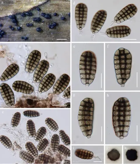
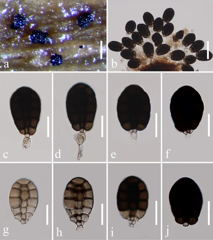
Canalisporium kenyense (MFLU 17-1086, reference specimen). a Colony on substrate. b–e Conidia with attached conidiogenous cells. f–j Conidia. Scale bars a = 200 µm, b = 50 µm, c–j = 20 µm
- Jenis
- Pelat ilmiah
- Kredit
- Penulis dan sumber artikel: Phookamsak, Rungtiwa; Hyde, Kevin D.; Jeewon, Rajesh; Bhat, D. Jayarama; Jones, E. B. Gareth; Maharachchikumbura, Sajeewa S. N.; Raspé, Olivier; Karunarathna, Samantha C.; Wanasinghe, Dhanushka N.; Hongsanan, Sinang; Doilom, Mingkwan; Tennakoon, Danushka S.; Machado, Alexandre R.; Firmino, André L.; Ghosh, Aniket; Karunarathna, Anuruddha; Mešić, Armin; Dutta, Arun Kumar; Thongbai, Benjarong; Devadatha, Bandarupalli; Norphanphoun, Chada; Senwanna, Chanokned; Wei, Deping; Pem, Dhandevi; Ackah, Frank Kwekucher; Wang, Gen-Nuo; Jiang, Hong-Bo; Madrid, Hugo; Lee, Hyang Burm; Goonasekara, Ishani D.; Manawasinghe, Ishara S.; Kušan, Ivana; Cano, Josep; Gené, Josepa; Li, Junfu; Das, Kanad; Acharya, Krishnendu; Raj, K. N. Anil; Latha, K. P. Deepna; Chethana, K. W. Thilini; He, Mao-Qiang; Dueñas, Margarita; Jadan, Margita; Martín, María P.; Samarakoon, Milan C.; Dayarathne, Monika C.; Raza, Mubashar; Park, Myung Soo; Telleria, M. Teresa; Chaiwan, Napalai; Matočec, Neven; de Silva, Nimali I.; Pereira, Olinto L.; Singh, Paras Nath; Manimohan, Patinjareveettil; Uniyal, Priyanka; Shang, Qiu-Ju; Bhatt, Rajendra P.; Perera, Rekhani H.; Alvarenga, Renato Lúcio Mendes; Nogal-Prata, Sandra; Singh, Sanjay K.; Vadthanarat, Santhiti; Oh, Seung-Yoon; Huang, Shi-Ke; Rana, Shiwali; Konta, Sirinapa; Paloi, Soumitra; Jayasiri, Subashini C.; Jeon, Sun Jeong; Mehmood, Tahir; Gibertoni, Tatiana Baptista; Nguyen, Thuong T. T.; Singh, Upendra; Thiyagaraja, Vinodhini; Sarma, V. Venkateswara; Dong, Wei; Yu, Xian-Dong; Lu, Yong-Zhong; Lim, Young Woon; Chen, Yun; Tkalčec, Zdenko; Zhang, Zhi-Feng; Luo, Zong-Long; Daranagama, Dinushani A.; Thambugala, Kasun M.; Tibpromma, Saowaluck; Camporesi, Erio; Bulgakov, Timur S.; Dissanayake, Asha J.; Senanayake, Indunil C.; Dai, Dong Qin; Tang, Li-Zhou; Khan, Sehroon; Zhang, Huang; Promputtha, Itthayakorn; Cai, Lei; Chomnunti, Putarak; Zhao, Rui-Lin; Lumyong, Saisamorn; Boonmee, Saranyaphat; Wen, Ting-Chi; Mortimer, Peter E.; Xu, Jianchu
- Lisensi
- CC BY 4.0
- Sumber media
- Buka entri sumber
Sumber profil
- [1] Fungal diversity notes 929–1035: taxonomic and phylogenetic contributions on genera and species of fungi — Canalisporium kenyense
- [2] Catalogue of Life Base2026-09-11 — Canalisporium kenyense
- [3] Dokumentasi visual — Canalisporium kenyense
Rujukan halaman ini
Fungi. Canalisporium kenyense. Rilis data fungi-2026-10-12-expansion-2. https://fungi-atlas.mgadib.chatgpt.site/jamur/canalisporium-kenyense/.
Status: impor sumber; pemeriksaan editorial per profil belum selesai. Untuk klaim biologis, gunakan rujukan pada bagian terkait dan periksa konteks sumber aslinya.