01 / Morfologi

Ciri dan struktur

Colonies on PDA at 25 °C after 3 d, white, reaching ca. 7–10 mm in diameter. Mycelia colourless, 3–4.5 μm wide. Primary conidiophores, colourless, unbranched and producing a single globose conidium with widening upwards; they offer a pronounced dimensional contrast with the mycelial filaments, extending to a length of 50–210 μm into the air, 3–25 μm wide. Primary conidia forcibly discharged, colourless, globose, measuring 15–45 μm in greatest length and 13–42 μm in total width, including a basal papilla 2–6 μm high and 5–17 μm wide. After discharging on to 2% water-agar, similar and smaller secondary conidia arise from primary conidia, two generations of multiple spherical units forming on the parent globose conidia Microconidia only formed from the second set, 5–12 × 9–10 μm. Capilliconidia formed readily from discharged microconidia, 16–24 × 5–6 μm. Chlamydospores formed within the substratum, colourless, globose to ellipsoidal, 13–40 × 15–45 μm.

Sebagian uraian ditampilkan dalam bahasa sumber; terjemahannya belum tersedia atau belum lolos pemeriksaan.

Rujukan: [1]

Pengukuran mengikuti objek, spesimen atau kultur, dan metode yang disebutkan sumber. Baca uraian asli untuk konteks lengkap.

02 / Kehidupan

Habitat dan peran ekologis

Colonies on PDA at 25 °C after 3 d, white, reaching ca. 7–10 mm in diameter. Mycelia colourless, 3–4.5 μm wide. Primary conidiophores, colourless, unbranched and producing a single globose conidium with widening upwards; they offer a pronounced dimensional contrast with the mycelial filaments, extending to a length of 50–210 μm into the air, 3–25 μm wide. Primary conidia forcibly discharged, colourless, globose, measuring 15–45 μm in greatest length and 13–42 μm in total width, including a basal papilla 2–6 μm high and 5–17 μm wide. After discharging on to 2% water-agar, similar and smaller secondary conidia arise from primary conidia, two generations of multiple spherical units forming on the parent globose conidia Microconidia only formed from the second set, 5–12 × 9–10 μm. Capilliconidia formed readily from discharged microconidia, 16–24 × 5–6 μm. Chlamydospores formed within the substratum, colourless, globose to ellipsoidal, 13–40 × 15–45 μm.

Sebagian uraian ditampilkan dalam bahasa sumber; terjemahannya belum tersedia atau belum lolos pemeriksaan.

Rujukan: [1]

Habitat atau konteks temuan
kultur yang didokumentasikan
Substrat
Informasi belum tersedia pada sumber rilis ini.
Peran atau konteks
Belum dikategorikan pada rilis ini.
Temukan hubungan serupa
03 / Siklus hidup

Reproduksi

Colonies on PDA at 25 °C after 3 d, white, reaching ca. 7–10 mm in diameter. Mycelia colourless, 3–4.5 μm wide. Primary conidiophores, colourless, unbranched and producing a single globose conidium with widening upwards; they offer a pronounced dimensional contrast with the mycelial filaments, extending to a length of 50–210 μm into the air, 3–25 μm wide. Primary conidia forcibly discharged, colourless, globose, measuring 15–45 μm in greatest length and 13–42 μm in total width, including a basal papilla 2–6 μm high and 5–17 μm wide. After discharging on to 2% water-agar, similar and smaller secondary conidia arise from primary conidia, two generations of multiple spherical units forming on the parent globose conidia Microconidia only formed from the second set, 5–12 × 9–10 μm.

Sebagian uraian ditampilkan dalam bahasa sumber; terjemahannya belum tersedia atau belum lolos pemeriksaan.

Rujukan: [1]

Sinonim menurut acuan taksonomi rilis: Conidiobolus adiaeretus
04 / Batas informasi

Catatan identitas dan cakupan

  • Nama diterima mengikuti Catalogue of Life Base2026-09-11; uraian mempertahankan konsep dan nama sumber aslinya.
  • Preserve original treatment localities, hosts and substrate scope. Materials examined describe documented specimens, not the full global range.
  • Impor dan terjemahan otomatis belum diperiksa editorial per profil. Struktur reproduktif yang disebutkan tidak membuktikan seluruh siklus hidup diketahui.
  • Pengukuran pada uraian mengikuti spesimen, kultur, dan metode sumber; angka tidak diperluas ke semua populasi.

Catatan distribusi dalam narasi mengikuti sumbernya. Dokumentasi spesimen atau foto mengikuti catatan sumber. Bukti wilayah Indonesia dan penilaian konservasi tidak disimpulkan dari gambar.

05 / Dokumentasi

Melihat dengan konteks

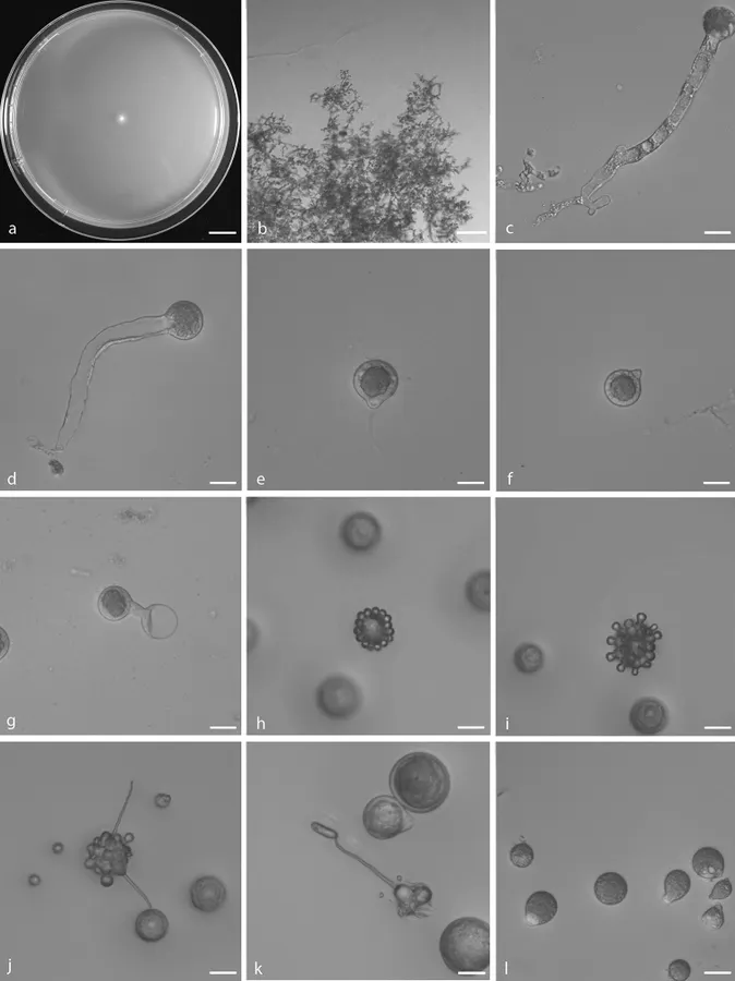
Capillidium adiaeretum a colony on PDA after 3 d at 25 °C b mycelia c, d primary conidiophores bearing primary conidia e, f primary conidia g Production of secondary conidia h first stage of forming microconidia i second stage of forming microconidia j, k ellipsoidal secondary conidia arising from slender conidiophores l chlamydospores. Scale bars: 10 mm (a); 100 μm (b); 20 μm (c–l).

Jenis
Pelat ilmiah
Kredit
Yong Nie, De-Shui Yu, Cheng-Fang Wang, Xiao-Yong Liu, Bo Huang
Lisensi
CC BY 4.0
Sumber media
Buka entri sumber

Gambar menunjukkan objek menurut identifikasi sumber, bukan verifikasi independen spesimen oleh Fungi. Tidak ada skala baru yang ditambahkan.

06 / Rujukan

Sumber profil

  1. [1] A taxonomic revision of the genus Conidiobolus (Ancylistaceae, Entomophthorales): four clades including three new genera — Capillidium adiaeretumBagian sumber: PMC7136305_section_17
    Tanggal penghimpunan sumber: 2026-10-12
    Teks sumber: CC BY 4.0 · Nie Y, Yu DS, Wang CF, Liu XY, Huang B.
  2. [2] Catalogue of Life Base2026-09-11 — Capillidium adiaeretumBagian sumber: ColDP NameUsage.tsv ID 9Z2HJ; accepted species; source 2073
    Tanggal penghimpunan sumber: 2026-10-12
  3. [3] Dokumentasi visual — Capillidium adiaeretumBagian sumber: Individual media record; identification follows the source, not independent determination
    Tanggal penghimpunan sumber: 2026-10-12
    Media sumber: CC BY 4.0 · Yong Nie, De-Shui Yu, Cheng-Fang Wang, Xiao-Yong Liu, Bo Huang

Adaptasi teks: CC BY 4.0. Nie Y, Yu DS, Wang CF, Liu XY, Huang B. — A taxonomic revision of the genus Conidiobolus (Ancylistaceae, Entomophthorales): four clades including three new genera — Capillidium adiaeretum

Uraian asli dan riwayat sumber

Riwayat atau halaman sumber

Ringkasan sumber

Colonies on PDA at 25 °C after 3 d, white, reaching ca. 7–10 mm in diameter. Mycelia colourless, 3–4.5 μm wide. Primary conidiophores, colourless, unbranched and producing a single globose conidium with widening upwards; they offer a pronounced dimensional contrast with the mycelial filaments, extending to a length of 50–210 μm into the air, 3–25 μm wide. Primary conidia forcibly discharged, colourless, globose, measuring 15–45 μm in greatest length and 13–42 μm in total width, including a basal papilla 2–6 μm high and 5–17 μm wide.

Revisi sumber

Morfologi sumber

Colonies on PDA at 25 °C after 3 d, white, reaching ca. 7–10 mm in diameter. Mycelia colourless, 3–4.5 μm wide. Primary conidiophores, colourless, unbranched and producing a single globose conidium with widening upwards; they offer a pronounced dimensional contrast with the mycelial filaments, extending to a length of 50–210 μm into the air, 3–25 μm wide. Primary conidia forcibly discharged, colourless, globose, measuring 15–45 μm in greatest length and 13–42 μm in total width, including a basal papilla 2–6 μm high and 5–17 μm wide. After discharging on to 2% water-agar, similar and smaller secondary conidia arise from primary conidia, two generations of multiple spherical units forming on the parent globose conidia Microconidia only formed from the second set, 5–12 × 9–10 μm. Capilliconidia formed readily from discharged microconidia, 16–24 × 5–6 μm. Chlamydospores formed within the substratum, colourless, globose to ellipsoidal, 13–40 × 15–45 μm.

Revisi sumber

Ekologi sumber

Colonies on PDA at 25 °C after 3 d, white, reaching ca. 7–10 mm in diameter. Mycelia colourless, 3–4.5 μm wide. Primary conidiophores, colourless, unbranched and producing a single globose conidium with widening upwards; they offer a pronounced dimensional contrast with the mycelial filaments, extending to a length of 50–210 μm into the air, 3–25 μm wide. Primary conidia forcibly discharged, colourless, globose, measuring 15–45 μm in greatest length and 13–42 μm in total width, including a basal papilla 2–6 μm high and 5–17 μm wide. After discharging on to 2% water-agar, similar and smaller secondary conidia arise from primary conidia, two generations of multiple spherical units forming on the parent globose conidia Microconidia only formed from the second set, 5–12 × 9–10 μm. Capilliconidia formed readily from discharged microconidia, 16–24 × 5–6 μm. Chlamydospores formed within the substratum, colourless, globose to ellipsoidal, 13–40 × 15–45 μm.

Revisi sumber

Struktur reproduksi sumber

Colonies on PDA at 25 °C after 3 d, white, reaching ca. 7–10 mm in diameter. Mycelia colourless, 3–4.5 μm wide. Primary conidiophores, colourless, unbranched and producing a single globose conidium with widening upwards; they offer a pronounced dimensional contrast with the mycelial filaments, extending to a length of 50–210 μm into the air, 3–25 μm wide. Primary conidia forcibly discharged, colourless, globose, measuring 15–45 μm in greatest length and 13–42 μm in total width, including a basal papilla 2–6 μm high and 5–17 μm wide. After discharging on to 2% water-agar, similar and smaller secondary conidia arise from primary conidia, two generations of multiple spherical units forming on the parent globose conidia Microconidia only formed from the second set, 5–12 × 9–10 μm.

Revisi sumber

Rujukan halaman ini

Fungi. Capillidium adiaeretum. Rilis data fungi-2026-10-12-expansion-2. https://fungi-atlas.mgadib.chatgpt.site/jamur/capillidium-adiaeretum/.

Status: impor sumber; pemeriksaan editorial per profil belum selesai. Untuk klaim biologis, gunakan rujukan pada bagian terkait dan periksa konteks sumber aslinya.