01 / Morfologi

Ciri dan struktur

Spores 6.5x4-6.5 (5.2x5.l)µm, Q= 1.01, globose to sub-globose, occasionally broadly pyriform, hyaline, inamyloid, not dextrinoid, acyanophilic, wall very thin, collapsing easily, apiculus usually short and distinct, occasionally extending gradually from the spore to give the pyriform spore outline, often with a distinct subterminal scar. Basidia 20-22 x 8 µm, subcylindrical to clavate, relatively short and stout, extending slightly at maturity; sterigmata (2)-4, 3-4 µm long, often scarred apically. Cheilocystidia and pleurocystidia absent. Trama fairly dense, somewhat bilateral with a central region of more or less parallel hyphae, 3-4 µm diam., lateral hyphae more interwoven, slightly divergent, merging into the narrow, filamentous subhymenium. Context of fairly densely packed hyphae, 5-6 µm diam., more parallel than interwoven and difficult to squash, merging into the loosely arranged pileipellis region with upturned hyphal endings responsible for the velutinous appearance and bundles of narrower hyphae for the villi. The pileus margin is heteromorphous, composed mainly of short, cylindrical to subclavate, flexuose to geniculate hyphal endings, some basidia and a number of more irregular cells basically narrow-subulate with a distinctive, long narrow neck. All hyphae have conspicuous clamp connections, and there was no indication of a gelatinisation.

Sebagian uraian ditampilkan dalam bahasa sumber; terjemahannya belum tersedia atau belum lolos pemeriksaan.

Rujukan: [1]

Pengukuran mengikuti objek, spesimen atau kultur, dan metode yang disebutkan sumber. Baca uraian asli untuk konteks lengkap.

02 / Kehidupan

Habitat dan peran ekologis

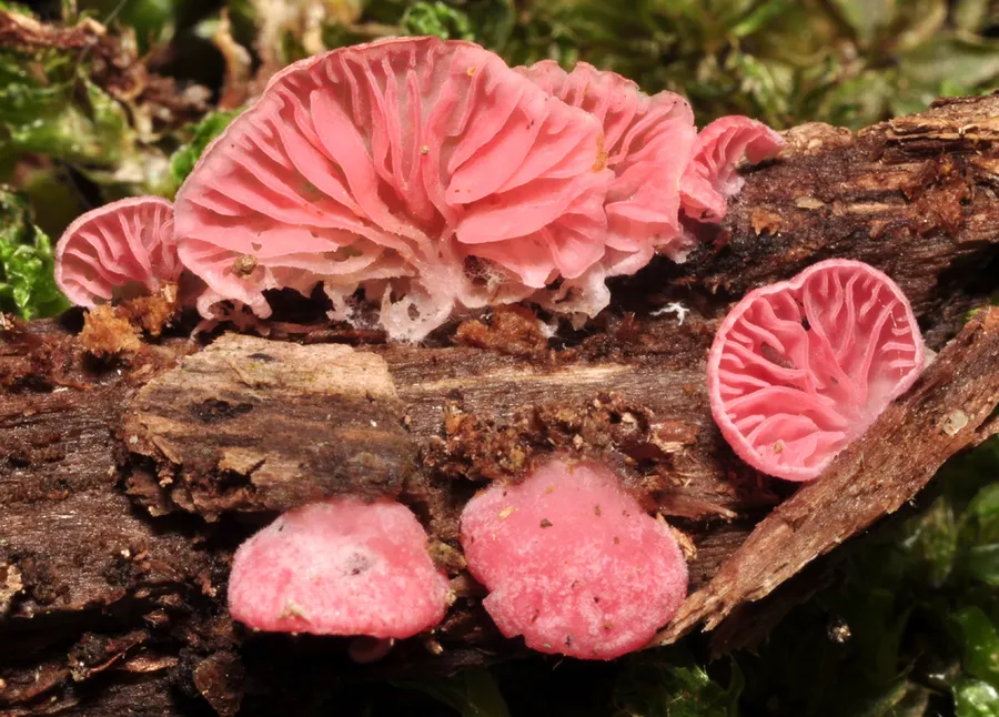
Gregarious on dead wood on the ground in Nothofagus forest. The name reflects the all-over pink colour of the basidiome Cheimonophyllum roseum was found quite commonly on dead twigs and branches of Nothofagus fusca and a N. menziesii in the Lewis Pass area. Its all-over shell-pink colour makes it quite distinctive as compared with other described species of the genus, which are all reported to have white basidiomes. It also has broader lamellae, without interveining, and lacks a stipe. As in C. candidissimum, no zone of cheilocystidia was found, but some awl-shaped cells were found on pileus margins of C. roseum. These are very similar to the awl-shaped cheilocystidia described by Horak (1968) and Pegler (1983) for C. candidissimum, and are also believed to be hyphal outgrowths from the trama or context, as described by Reijnders & Stalpers (1992).

Sebagian uraian ditampilkan dalam bahasa sumber; terjemahannya belum tersedia atau belum lolos pemeriksaan.

Rujukan: [1]

Habitat atau konteks temuan
hutan
Substrat
kayu; tanah
Peran atau konteks
Belum dikategorikan pada rilis ini.
Temukan hubungan serupa
03 / Siklus hidup

Reproduksi

Spores 6.5x4-6.5 (5.2x5.l)µm, Q= 1.01, globose to sub-globose, occasionally broadly pyriform, hyaline, inamyloid, not dextrinoid, acyanophilic, wall very thin, collapsing easily, apiculus usually short and distinct, occasionally extending gradually from the spore to give the pyriform spore outline, often with a distinct subterminal scar. Basidia 20-22 x 8 µm, subcylindrical to clavate, relatively short and stout, extending slightly at maturity; sterigmata (2)-4, 3-4 µm long, often scarred apically. Cheilocystidia and pleurocystidia absent. Trama fairly dense, somewhat bilateral with a central region of more or less parallel hyphae, 3-4 µm diam., lateral hyphae more interwoven, slightly divergent, merging into the narrow, filamentous subhymenium.

Sebagian uraian ditampilkan dalam bahasa sumber; terjemahannya belum tersedia atau belum lolos pemeriksaan.

Rujukan: [1]

04 / Batas informasi

Catatan identitas dan cakupan

  • Nama diterima mengikuti Catalogue of Life Base2026-09-11; uraian mempertahankan konsep dan nama sumber aslinya.
  • Historical description as cited by BiotaNZ. Preserve explicit New Zealand/locality, host and specimen qualifiers. Distribution is the cited source scope, not a current global range.
  • Impor dan terjemahan otomatis belum diperiksa editorial per profil. Struktur reproduktif yang disebutkan tidak membuktikan seluruh siklus hidup diketahui.
  • Pengukuran pada uraian mengikuti spesimen, kultur, dan metode sumber; angka tidak diperluas ke semua populasi.

Catatan distribusi dalam narasi mengikuti sumbernya. Dokumentasi spesimen atau foto mengikuti catatan sumber. Bukti wilayah Indonesia dan penilaian konservasi tidak disimpulkan dari gambar.

05 / Dokumentasi

Melihat dengan konteks

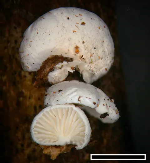
Dokumentasi Cheimonophyllum roseum menurut asosiasi foto pada record takson iNaturalist. Kredit: Jerry Cooper. Identitas mengikuti sumber; bukan penentuan spesimen independen.

Jenis
Fotografi
Kredit
(c) Jerry Cooper, some rights reserved (CC BY), uploaded by Jerry Cooper
Lisensi
CC BY 4.0
Sumber media
Buka entri sumber

Gambar menunjukkan objek menurut identifikasi sumber, bukan verifikasi independen spesimen oleh Fungi. Tidak ada skala baru yang ditambahkan.

06 / Rujukan

Sumber profil

  1. [1] Segedin, B.P. 1994: Studies in the Agaricales of New Zealand: new records and new species of the genera Cheimonophyllum, Mniopetalum, and Anthracophyllum (Tricholomataceae, Collybieae). New Zealand Journal of Botany 32: 61-72. — Cheimonophyllum roseumBagian sumber: biota:1cb0faea-36b9-11d5-9548-00d0592d548c:1cb18257-36b9-11d5-9548-00d0592d548c
    Tanggal penghimpunan sumber: 2026-10-12
    Teks sumber: CC BY 4.0 · Segedin, B.P. 1994: Studies in the Agaricales of New Zealand: new records and new species of the genera Cheimonophyllum, Mniopetalum, and Anthracophyllum (Tricholomataceae, Collybieae). New Zealand Journal of Botany 32: 61-72.
  2. [2] Catalogue of Life Base2026-09-11 — Cheimonophyllum roseumBagian sumber: ColDP NameUsage.tsv ID TT97; accepted species; source 2073
    Tanggal penghimpunan sumber: 2026-10-12
  3. [3] Dokumentasi visual — Cheimonophyllum roseumBagian sumber: Individual media record; identification follows the source, not independent determination
    Tanggal penghimpunan sumber: 2026-10-12
    Media sumber: CC BY 4.0 · (c) Jerry Cooper, some rights reserved (CC BY), uploaded by Jerry Cooper

Adaptasi teks: CC BY 4.0. Segedin, B.P. 1994: Studies in the Agaricales of New Zealand: new records and new species of the genera Cheimonophyllum, Mniopetalum, and Anthracophyllum (Tricholomataceae, Collybieae). New Zealand Journal of Botany 32: 61-72. — Segedin, B.P. 1994: Studies in the Agaricales of New Zealand: new records and new species of the genera Cheimonophyllum, Mniopetalum, and Anthracophyllum (Tricholomataceae, Collybieae). New Zealand Journal of Botany 32: 61-72. — Cheimonophyllum roseum

Uraian asli dan riwayat sumber

Riwayat atau halaman sumber

Ringkasan sumber

Spores 6.5x4-6.5 (5.2x5.l)µm, Q= 1.01, globose to sub-globose, occasionally broadly pyriform, hyaline, inamyloid, not dextrinoid, acyanophilic, wall very thin, collapsing easily, apiculus usually short and distinct, occasionally extending gradually from the spore to give the pyriform spore outline, often with a distinct subterminal scar. Basidia 20-22 x 8 µm, subcylindrical to clavate, relatively short and stout, extending slightly at maturity; sterigmata (2)-4, 3-4 µm long, often scarred apically.

Revisi sumber

Morfologi sumber

Spores 6.5x4-6.5 (5.2x5.l)µm, Q= 1.01, globose to sub-globose, occasionally broadly pyriform, hyaline, inamyloid, not dextrinoid, acyanophilic, wall very thin, collapsing easily, apiculus usually short and distinct, occasionally extending gradually from the spore to give the pyriform spore outline, often with a distinct subterminal scar. Basidia 20-22 x 8 µm, subcylindrical to clavate, relatively short and stout, extending slightly at maturity; sterigmata (2)-4, 3-4 µm long, often scarred apically. Cheilocystidia and pleurocystidia absent. Trama fairly dense, somewhat bilateral with a central region of more or less parallel hyphae, 3-4 µm diam., lateral hyphae more interwoven, slightly divergent, merging into the narrow, filamentous subhymenium. Context of fairly densely packed hyphae, 5-6 µm diam., more parallel than interwoven and difficult to squash, merging into the loosely arranged pileipellis region with upturned hyphal endings responsible for the velutinous appearance and bundles of narrower hyphae for the villi. The pileus margin is heteromorphous, composed mainly of short, cylindrical to subclavate, flexuose to geniculate hyphal endings, some basidia and a number of more irregular cells basically narrow-subulate with a distinctive, long narrow neck. All hyphae have conspicuous clamp connections, and there was no indication of a gelatinisation.

Revisi sumber

Ekologi sumber

Gregarious on dead wood on the ground in Nothofagus forest. The name reflects the all-over pink colour of the basidiome Cheimonophyllum roseum was found quite commonly on dead twigs and branches of Nothofagus fusca and a N. menziesii in the Lewis Pass area. Its all-over shell-pink colour makes it quite distinctive as compared with other described species of the genus, which are all reported to have white basidiomes. It also has broader lamellae, without interveining, and lacks a stipe. As in C. candidissimum, no zone of cheilocystidia was found, but some awl-shaped cells were found on pileus margins of C. roseum. These are very similar to the awl-shaped cheilocystidia described by Horak (1968) and Pegler (1983) for C. candidissimum, and are also believed to be hyphal outgrowths from the trama or context, as described by Reijnders & Stalpers (1992).

Revisi sumber

Struktur reproduksi sumber

Spores 6.5x4-6.5 (5.2x5.l)µm, Q= 1.01, globose to sub-globose, occasionally broadly pyriform, hyaline, inamyloid, not dextrinoid, acyanophilic, wall very thin, collapsing easily, apiculus usually short and distinct, occasionally extending gradually from the spore to give the pyriform spore outline, often with a distinct subterminal scar. Basidia 20-22 x 8 µm, subcylindrical to clavate, relatively short and stout, extending slightly at maturity; sterigmata (2)-4, 3-4 µm long, often scarred apically. Cheilocystidia and pleurocystidia absent. Trama fairly dense, somewhat bilateral with a central region of more or less parallel hyphae, 3-4 µm diam., lateral hyphae more interwoven, slightly divergent, merging into the narrow, filamentous subhymenium.

Revisi sumber

Rujukan halaman ini

Fungi. Cheimonophyllum roseum. Rilis data fungi-2026-10-12-expansion-2. https://fungi-atlas.mgadib.chatgpt.site/jamur/cheimonophyllum-roseum/.

Status: impor sumber; pemeriksaan editorial per profil belum selesai. Untuk klaim biologis, gunakan rujukan pada bagian terkait dan periksa konteks sumber aslinya.