Ciri dan struktur
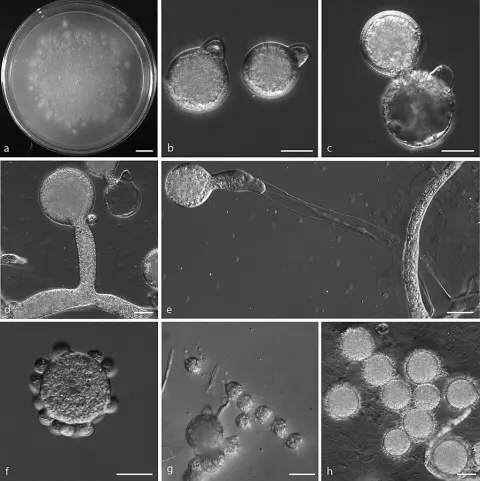
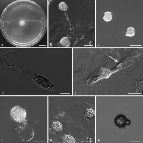
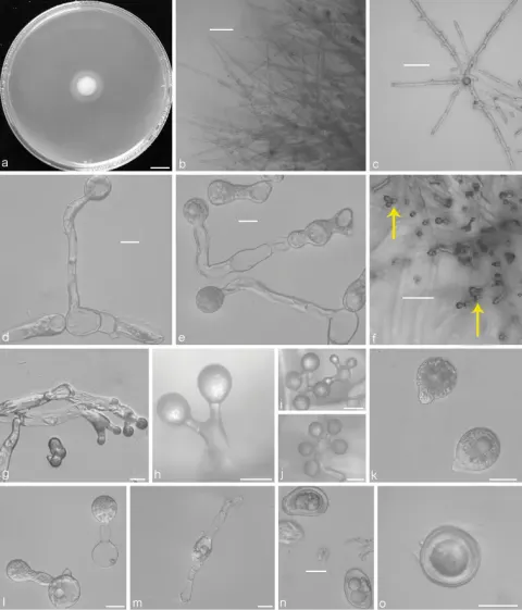
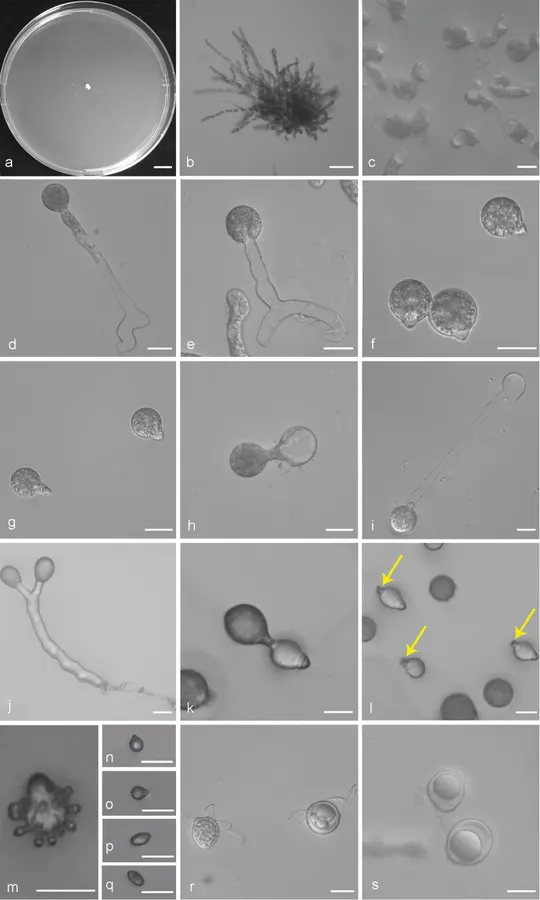
Colonies on PDA at 21 °C for 3 d, opaque, white, reaching ca. 2 mm in diameter, with many small colonies around the periphery due to discharged conidia. Mycelia colourless, 8–11 μm wide, rarely branched and non-septate when young, often septate and distended to a width of 10–27 μm after 5 d. Primary conidiophores arising from the hyphal segments, colourless, 38–254 × 7.5–12 μm, unbranched and producing a single globose conidium, without widening upwards near the tip. Primary conidia forcibly discharged, globose to subglobose, 2–40 × 2–33 μm, with a papilla more or less tapering and pointed, 7–11 μm wide at the base, 3–12 μm long. Secondary conidiophores arising from the primary conidia, often branched almost at the tip, forming two short stipes each bearing a secondary conidium. Secondary conidia similar to, but smaller than the primary ones, mostly forcibly discharged, occasionally falling off and leaving a relic of the secondary conidiophores. On 2 % water agar, microconidia produced readily, globose to ellipsoidal, 7–12 × 6–9 μm. Zygospores homothallic, usually formed between adjacent segments of the same hypha after an incubation of 5–7 d at 21 °C on PDA, smooth, mostly globose, 25–40 μm in diameter, with a 1.5–3 μm thick wall.
Habitat dan peran ekologis
Colonies on PDA at 21 °C for 3 d, opaque, white, reaching ca. 2 mm in diameter, with many small colonies around the periphery due to discharged conidia. Mycelia colourless, 8–11 μm wide, rarely branched and non-septate when young, often septate and distended to a width of 10–27 μm after 5 d. Primary conidiophores arising from the hyphal segments, colourless, 38–254 × 7.5–12 μm, unbranched and producing a single globose conidium, without widening upwards near the tip. Primary conidia forcibly discharged, globose to subglobose, 2–40 × 2–33 μm, with a papilla more or less tapering and pointed, 7–11 μm wide at the base, 3–12 μm long. Secondary conidiophores arising from the primary conidia, often branched almost at the tip, forming two short stipes each bearing a secondary conidium. Secondary conidia similar to, but smaller than the primary ones, mostly forcibly discharged, occasionally falling off and leaving a relic of the secondary conidiophores. On 2 % water agar, microconidia produced readily, globose to ellipsoidal, 7–12 × 6–9 μm. Zygospores homothallic, usually formed between adjacent segments of the same hypha after an incubation of 5–7 d at 21 °C on PDA, smooth, mostly globose, 25–40 μm in diameter, with a 1.5–3 μm thick wall.
- Habitat atau konteks temuan
- kultur yang didokumentasikan
- Substrat
- Informasi belum tersedia pada sumber rilis ini.
- Peran atau konteks
- Belum dikategorikan pada rilis ini.
Reproduksi
Colonies on PDA at 21 °C for 3 d, opaque, white, reaching ca. 2 mm in diameter, with many small colonies around the periphery due to discharged conidia. Mycelia colourless, 8–11 μm wide, rarely branched and non-septate when young, often septate and distended to a width of 10–27 μm after 5 d. Primary conidiophores arising from the hyphal segments, colourless, 38–254 × 7.5–12 μm, unbranched and producing a single globose conidium, without widening upwards near the tip. Primary conidia forcibly discharged, globose to subglobose, 2–40 × 2–33 μm, with a papilla more or less tapering and pointed, 7–11 μm wide at the base, 3–12 μm long. Secondary conidiophores arising from the primary conidia, often branched almost at the tip, forming two short stipes each bearing a secondary conidium.
Catatan identitas dan cakupan
- Nama diterima mengikuti Catalogue of Life Base2026-09-11; uraian mempertahankan konsep dan nama sumber aslinya.
- Preserve original treatment localities, hosts and substrate scope. Materials examined describe documented specimens, not the full global range.
- Impor dan terjemahan otomatis belum diperiksa editorial per profil. Struktur reproduktif yang disebutkan tidak membuktikan seluruh siklus hidup diketahui.
- Pengukuran pada uraian mengikuti spesimen, kultur, dan metode sumber; angka tidak diperluas ke semua populasi.
Catatan distribusi dalam narasi mengikuti sumbernya. Dokumentasi spesimen atau foto mengikuti catatan sumber. Bukti wilayah Indonesia dan penilaian konservasi tidak disimpulkan dari gambar.
Melihat dengan konteks
Conidiobolus bifurcatus sp. nov. a Colony on PDA after 3 d at 21 °C b mycelium c septate mycelium and distended segments d, e primary conidiophores bearing primary conidia f, g primary conidia h, i a single secondary conidium produced from primary conidia j two secondary conidia arising from a branched conidiophore k secondary conidia falling from primary conidia l the relic of secondary conidiophores on secondary conidia (arrows) m microconidia arising from a conidium n, o globose microconidia p, q ellipsoidal microconidia r zygospores formed between adjacent segments of the same hypha s zygospores. Scale bars: 10 mm (a); 100 μm (b); 20 μm (c–s).
- Jenis
- Pelat ilmiah
- Kredit
- Yong Nie, Yue Cai, Yang Gao, De-Shui Yu, Zi-Min Wang, Xiao-Yong Liu, Bo Huang
- Lisensi
- CC BY 4.0
- Sumber media
- Buka entri sumber
Sumber profil
- [1] Three new species of Conidiobolus sensu stricto from plant debris in eastern China — Conidiobolus bifurcatus
- [2] Catalogue of Life Base2026-09-11 — Conidiobolus bifurcatus
- [3] Dokumentasi visual — Conidiobolus bifurcatus
Rujukan halaman ini
Fungi. Conidiobolus bifurcatus. Rilis data fungi-2026-10-12-expansion-2. https://fungi-atlas.mgadib.chatgpt.site/jamur/conidiobolus-bifurcatus/.
Status: impor sumber; pemeriksaan editorial per profil belum selesai. Untuk klaim biologis, gunakan rujukan pada bagian terkait dan periksa konteks sumber aslinya.