01 / Morfologi

Ciri dan struktur

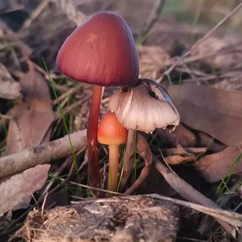
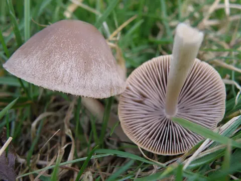
Pileus <35 mm, convex-umbonate to plano-convex but retaining umbo, yellow-brown or tawny brown, drying paler (yellowish), disc rather darker, moist, matt, radially wrinkled about umbo; margin radially striate, splitting. Stipe 35-40 x 3 mm, equal but for abruptly bulbous base, concolorous or paler than pileus, hollow. Gills adnexed, yellow-brown, crowded. Flesh extremely thin in pileus, whitish, distinct from the easily removable stipe, fibrous, very brittle in stipe. Basidiospores brownish yellow-ochre in mass, 5.5-7(8.5) x 3.5-4.5 µm, elongate-elliptic in face-view, perhaps slightly amygdaliform in side-view, smooth, non-amyloid, slightly thick-walled, ochraceous in water; germ-pore absent. Basidia 4-spored, hyaline, clavate-pedicellate. Hymenophoral trama subparallel. Cheilocystidia lecythiform, 16.5-21.5 x 6-7 µm, capitulum 3.5-4 µm, some with brownish plasmatic pigment in venter in KOH. Pileipellis a palisadoderm of pyriform to ellipsoid-pedicellate cells some with slightly brownish encrustations, 10-15 µm broad, some with plasmatic pigment, hair-like cells absent; pilocystidia detached, either long encrusted hyphae with clamp-connections (17.5-32 x (2.5-) 4.5-6 µm or short chains arising from ± vesiculose cells c. 12 µm broad, usually with swollen capitulum at apex and often containing brown plasmatic pigment. Pileus trama of hyphae with yellow contents in KOH. Stipitipellis of parallel to subparallel cells producing lageniform to cylindric caulocystidia intermixed with long flexuous hair-like cells <60 µm. Clamp-connections present in pileus trama, pileipellis and stipitipellis.

Sebagian uraian ditampilkan dalam bahasa sumber; terjemahannya belum tersedia atau belum lolos pemeriksaan.

Rujukan: [1]

Pengukuran mengikuti objek, spesimen atau kultur, dan metode yang disebutkan sumber. Baca uraian asli untuk konteks lengkap.

02 / Kehidupan

Habitat dan peran ekologis

On soil and in litter, under tawa (Beilschmiedia tawa Benth. & Hook. f.), Cyathea, Hedycarya, Schefflera and Alseuosmia, Mt Kaitarakihi track, Coromandel, 8 v 1977, Taylor 1055. The pilocystidia are distinctive and possibly represent remnants of the original blematogen as described for C. rickeniana (Singer) P D Orton and C. mesospora (Kuhner ex) Kuhner & Watling (Watling, 1975). Similar structures have not been found previously in C. piloselloides or C. pilosella (Pers.: Fr.) Kuhner and may indicate the New Zealand material represents an independent yet closely related taxon. Strong developments of pilocystidia are found in C. horakii Walling Taylor (q.v.) and in C. dumetorum (Vel.) Svreck The fungus is easily recognised by the small basidiospores lacking a germ-pore, strongly developed often-coloured pilocystidia and lageniform to cylindric caulocystidia mixed with hair-like cystidia. For these latter cells Walling (1964 et seq.) has erroneously used the term pilocystidia referring to the dermatocystidia of the pileus as pileocystidia. Patrick Barrows (1979) are correct in pointing out that pilocystidia a term introduced by Buller is derived from Greek where pile- refers to head and not hair (Latin).

Sebagian uraian ditampilkan dalam bahasa sumber; terjemahannya belum tersedia atau belum lolos pemeriksaan.

Rujukan: [1]

Habitat atau konteks temuan
Informasi belum tersedia pada sumber rilis ini.
Substrat
tanah
Peran atau konteks
Belum dikategorikan pada rilis ini.
Temukan hubungan serupa
03 / Siklus hidup

Reproduksi

Pileus <35 mm, convex-umbonate to plano-convex but retaining umbo, yellow-brown or tawny brown, drying paler (yellowish), disc rather darker, moist, matt, radially wrinkled about umbo; margin radially striate, splitting. Stipe 35-40 x 3 mm, equal but for abruptly bulbous base, concolorous or paler than pileus, hollow. Gills adnexed, yellow-brown, crowded. Flesh extremely thin in pileus, whitish, distinct from the easily removable stipe, fibrous, very brittle in stipe. Basidiospores brownish yellow-ochre in mass, 5.5-7(8.5) x 3.5-4.5 µm, elongate-elliptic in face-view, perhaps slightly amygdaliform in side-view, smooth, non-amyloid, slightly thick-walled, ochraceous in water; germ-pore absent.

Sebagian uraian ditampilkan dalam bahasa sumber; terjemahannya belum tersedia atau belum lolos pemeriksaan.

Rujukan: [1]

Sinonim menurut acuan taksonomi rilis: Agaricus pilosellus; Agaricus tener pilosellus; Conocybe pilosella var. brunneonigra; Conocybe piloselloides; Galera pilosella; Galera tenera var. pilosella; Galerula pilosella
04 / Batas informasi

Catatan identitas dan cakupan

  • Nama diterima mengikuti Catalogue of Life Base2026-09-11; uraian mempertahankan konsep dan nama sumber aslinya.
  • Historical description as cited by BiotaNZ. Preserve explicit New Zealand/locality, host and specimen qualifiers. Distribution is the cited source scope, not a current global range.
  • Impor dan terjemahan otomatis belum diperiksa editorial per profil. Struktur reproduktif yang disebutkan tidak membuktikan seluruh siklus hidup diketahui.
  • Pengukuran pada uraian mengikuti spesimen, kultur, dan metode sumber; angka tidak diperluas ke semua populasi.

Catatan distribusi dalam narasi mengikuti sumbernya. Dokumentasi spesimen atau foto mengikuti catatan sumber. Bukti wilayah Indonesia dan penilaian konservasi tidak disimpulkan dari gambar.

05 / Dokumentasi

Melihat dengan konteks

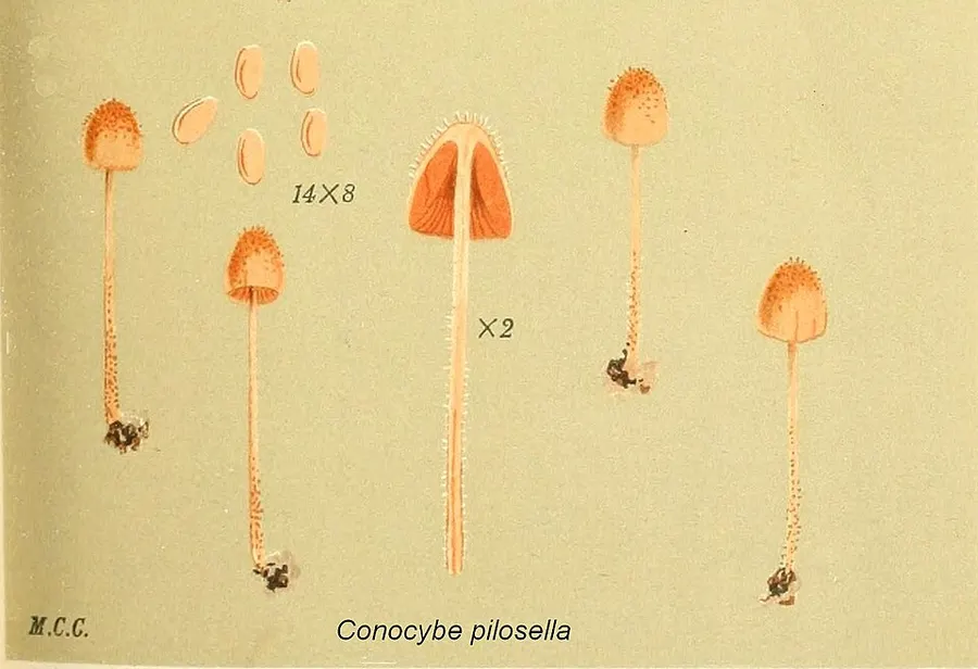
Dokumentasi Conocybe pilosella dari Wikimedia Commons. Conocybe pilosella Identifikasi mengikuti record berkas sumber.

Jenis
Fotografi
Kredit
author: Mordecai Cubitt Cooke, correct:Jerzy Opioła
Sumber media
Buka entri sumber

Gambar menunjukkan objek menurut identifikasi sumber, bukan verifikasi independen spesimen oleh Fungi. Tidak ada skala baru yang ditambahkan.

06 / Rujukan

Sumber profil

  1. [1] Watling, R.; Taylor, G.M. 1987: Observations on the Bolbitiaceae: 27. Preliminary account of the Bolbitiaceae of New Zealand. Bibliotheca Mycologica. 117. — Conocybe piloselloidesBagian sumber: biota:1cb0fec0-36b9-11d5-9548-00d0592d548c:1cb1b0a8-36b9-11d5-9548-00d0592d548c
    Tanggal penghimpunan sumber: 2026-10-12
    Teks sumber: CC BY 4.0 · Watling, R.; Taylor, G.M. 1987: Observations on the Bolbitiaceae: 27. Preliminary account of the Bolbitiaceae of New Zealand. Bibliotheca Mycologica. 117.
  2. [2] Catalogue of Life Base2026-09-11 — Conocybe pilosellaBagian sumber: ColDP NameUsage.tsv ID XQW9; accepted species; source 2073
    Tanggal penghimpunan sumber: 2026-10-12
  3. [3] Dokumentasi visual — Conocybe pilosellaBagian sumber: Individual media record; identification follows the source, not independent determination
    Tanggal penghimpunan sumber: 2026-10-12
    Media sumber: Public domain · author: Mordecai Cubitt Cooke, correct:Jerzy Opioła

Adaptasi teks: CC BY 4.0. Watling, R.; Taylor, G.M. 1987: Observations on the Bolbitiaceae: 27. Preliminary account of the Bolbitiaceae of New Zealand. Bibliotheca Mycologica. 117. — Watling, R.; Taylor, G.M. 1987: Observations on the Bolbitiaceae: 27. Preliminary account of the Bolbitiaceae of New Zealand. Bibliotheca Mycologica. 117. — Conocybe piloselloides

Uraian asli dan riwayat sumber

Riwayat atau halaman sumber

Ringkasan sumber

Pileus <35 mm, convex-umbonate to plano-convex but retaining umbo, yellow-brown or tawny brown, drying paler (yellowish), disc rather darker, moist, matt, radially wrinkled about umbo; margin radially striate, splitting. Stipe 35-40 x 3 mm, equal but for abruptly bulbous base, concolorous or paler than pileus, hollow. Gills adnexed, yellow-brown, crowded. Flesh extremely thin in pileus, whitish, distinct from the easily removable stipe, fibrous, very brittle in stipe. Basidiospores brownish yellow-ochre in mass, 5.5-7(8.5) x 3.5-4.5 µm, elongate-elliptic in face-view, perhaps slightly amygdaliform in side-view, smooth, non-amyloid, slightly thick-walled, ochraceous in water; germ-pore absent.

Revisi sumber

Morfologi sumber

Pileus <35 mm, convex-umbonate to plano-convex but retaining umbo, yellow-brown or tawny brown, drying paler (yellowish), disc rather darker, moist, matt, radially wrinkled about umbo; margin radially striate, splitting. Stipe 35-40 x 3 mm, equal but for abruptly bulbous base, concolorous or paler than pileus, hollow. Gills adnexed, yellow-brown, crowded. Flesh extremely thin in pileus, whitish, distinct from the easily removable stipe, fibrous, very brittle in stipe. Basidiospores brownish yellow-ochre in mass, 5.5-7(8.5) x 3.5-4.5 µm, elongate-elliptic in face-view, perhaps slightly amygdaliform in side-view, smooth, non-amyloid, slightly thick-walled, ochraceous in water; germ-pore absent. Basidia 4-spored, hyaline, clavate-pedicellate. Hymenophoral trama subparallel. Cheilocystidia lecythiform, 16.5-21.5 x 6-7 µm, capitulum 3.5-4 µm, some with brownish plasmatic pigment in venter in KOH. Pileipellis a palisadoderm of pyriform to ellipsoid-pedicellate cells some with slightly brownish encrustations, 10-15 µm broad, some with plasmatic pigment, hair-like cells absent; pilocystidia detached, either long encrusted hyphae with clamp-connections (17.5-32 x (2.5-) 4.5-6 µm or short chains arising from ± vesiculose cells c. 12 µm broad, usually with swollen capitulum at apex and often containing brown plasmatic pigment. Pileus trama of hyphae with yellow contents in KOH. Stipitipellis of parallel to subparallel cells producing lageniform to cylindric caulocystidia intermixed with long flexuous hair-like cells <60 µm. Clamp-connections present in pileus trama, pileipellis and stipitipellis.

Revisi sumber

Ekologi sumber

On soil and in litter, under tawa (Beilschmiedia tawa Benth. & Hook. f.), Cyathea, Hedycarya, Schefflera and Alseuosmia, Mt Kaitarakihi track, Coromandel, 8 v 1977, Taylor 1055. The pilocystidia are distinctive and possibly represent remnants of the original blematogen as described for C. rickeniana (Singer) P D Orton and C. mesospora (Kuhner ex) Kuhner & Watling (Watling, 1975). Similar structures have not been found previously in C. piloselloides or C. pilosella (Pers.: Fr.) Kuhner and may indicate the New Zealand material represents an independent yet closely related taxon. Strong developments of pilocystidia are found in C. horakii Walling Taylor (q.v.) and in C. dumetorum (Vel.) Svreck The fungus is easily recognised by the small basidiospores lacking a germ-pore, strongly developed often-coloured pilocystidia and lageniform to cylindric caulocystidia mixed with hair-like cystidia. For these latter cells Walling (1964 et seq.) has erroneously used the term pilocystidia referring to the dermatocystidia of the pileus as pileocystidia. Patrick Barrows (1979) are correct in pointing out that pilocystidia a term introduced by Buller is derived from Greek where pile- refers to head and not hair (Latin).

Revisi sumber

Struktur reproduksi sumber

Pileus <35 mm, convex-umbonate to plano-convex but retaining umbo, yellow-brown or tawny brown, drying paler (yellowish), disc rather darker, moist, matt, radially wrinkled about umbo; margin radially striate, splitting. Stipe 35-40 x 3 mm, equal but for abruptly bulbous base, concolorous or paler than pileus, hollow. Gills adnexed, yellow-brown, crowded. Flesh extremely thin in pileus, whitish, distinct from the easily removable stipe, fibrous, very brittle in stipe. Basidiospores brownish yellow-ochre in mass, 5.5-7(8.5) x 3.5-4.5 µm, elongate-elliptic in face-view, perhaps slightly amygdaliform in side-view, smooth, non-amyloid, slightly thick-walled, ochraceous in water; germ-pore absent.

Revisi sumber

Rujukan halaman ini

Fungi. Conocybe pilosella. Rilis data fungi-2026-10-12-expansion-2. https://fungi-atlas.mgadib.chatgpt.site/jamur/conocybe-pilosella/.

Status: impor sumber; pemeriksaan editorial per profil belum selesai. Untuk klaim biologis, gunakan rujukan pada bagian terkait dan periksa konteks sumber aslinya.