01 / Morfologi

Ciri dan struktur

Sexual morph: not observed. Asexual morph: Conidiomata pycnidial, immersed in bark, erumpent, ostiolated, with multiple irregularly arranged circular or ovoid locules, 1,050–1,400 μm in diam. Conceptacle absent, ostiole conspicuous, circular, dark gray, at the same level as the disc. Conidiophores semimacronematous, hyaline, filamentous, mainly unbranched or branched at base, elongated, smooth, thin-walled, (16.0–)17.7–22.2(–23.5) μm. Conidiogenous cells enteroblastic, polyphialidic. Conidia abundant, single, hyaline, aseptate, curved, allantoid, thin-walled (5.0–)5.4–6.1(–6.5) × (1.0–)1.2–1.5 μm. Colonies on MEA initially white, becoming beige with dense aerial mycelium, slow-growing (17 mm in diameter) after 7 days of incubation. Hyphae hyaline, smooth, branched, and septate.

Sebagian uraian ditampilkan dalam bahasa sumber; terjemahannya belum tersedia atau belum lolos pemeriksaan.

Rujukan: [1]

Pengukuran mengikuti objek, spesimen atau kultur, dan metode yang disebutkan sumber. Baca uraian asli untuk konteks lengkap.

02 / Kehidupan

Habitat dan peran ekologis

Morphologically, two isolates of C. piceae are very similar, but EI-19(A) has slightly longer conidiophores and conidia than CFCC 52841. Originally, C. piceae was described as a pathogen associated with the canker disease of Picea crassifolia in China (Pan et al. 2018). But the species was isolated from different hosts (including non-conifers) in other parts of Canada based on ITS sequence data (ON352565, PQ671332, PQ671333, PQ666762) available in GenBank.

Sebagian uraian ditampilkan dalam bahasa sumber; terjemahannya belum tersedia atau belum lolos pemeriksaan.

Rujukan: [1]

Habitat atau konteks temuan
Informasi belum tersedia pada sumber rilis ini.
Substrat
Informasi belum tersedia pada sumber rilis ini.
Peran atau konteks
Belum dikategorikan pada rilis ini.
Temukan hubungan serupa
03 / Siklus hidup

Reproduksi

Sexual morph: not observed. Asexual morph: Conidiomata pycnidial, immersed in bark, erumpent, ostiolated, with multiple irregularly arranged circular or ovoid locules, 1,050–1,400 μm in diam. Conceptacle absent, ostiole conspicuous, circular, dark gray, at the same level as the disc. Conidiophores semimacronematous, hyaline, filamentous, mainly unbranched or branched at base, elongated, smooth, thin-walled, (16.0–)17.7–22.2(–23.5) μm. Conidiogenous cells enteroblastic, polyphialidic. Conidia abundant, single, hyaline, aseptate, curved, allantoid, thin-walled (5.0–)5.4–6.1(–6.5) × (1.0–)1.2–1.5 μm.

Sebagian uraian ditampilkan dalam bahasa sumber; terjemahannya belum tersedia atau belum lolos pemeriksaan.

Rujukan: [1]

04 / Batas informasi

Catatan identitas dan cakupan

  • Nama diterima mengikuti Catalogue of Life Base2026-09-11; uraian mempertahankan konsep dan nama sumber aslinya.
  • Preserve original treatment localities, hosts and substrate scope. Materials examined describe documented specimens, not the full global range.
  • Impor dan terjemahan otomatis belum diperiksa editorial per profil. Struktur reproduktif yang disebutkan tidak membuktikan seluruh siklus hidup diketahui.
  • Pengukuran pada uraian mengikuti spesimen, kultur, dan metode sumber; angka tidak diperluas ke semua populasi.

Catatan distribusi dalam narasi mengikuti sumbernya. Dokumentasi spesimen atau foto mengikuti catatan sumber. Bukti wilayah Indonesia dan penilaian konservasi tidak disimpulkan dari gambar.

05 / Dokumentasi

Melihat dengan konteks

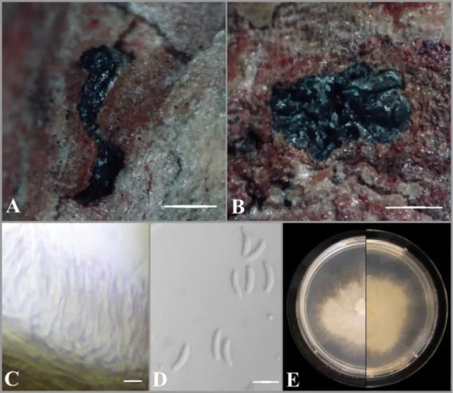
Asexual morph of Cytospora piceae A habit of conidiomata on twig of P. glauca B transverse section of conidioma C conidiophores and conidiogenous cells D conidia E adverse and reverse view of seven-day-old culture on MEA. Scale bars: 1 mm (A, B); 5 μm (C, D).

Jenis
Pelat ilmiah
Kredit
Evgeny Ilyukhin, Yanpeng Chen, Svetlana Markovskaja, Ashwag Shami, Sajeewa S. N. Maharachchikumbura
Lisensi
CC BY 4.0
Sumber media
Buka entri sumber

Gambar menunjukkan objek menurut identifikasi sumber, bukan verifikasi independen spesimen oleh Fungi. Tidak ada skala baru yang ditambahkan.

06 / Rujukan

Sumber profil

  1. [1] Comprehensive genome analysis of two Cytospora (Cytosporaceae, Diaporthales) species associated with canker disease of spruce: C. piceae and C. piceicola sp. nov. — Cytospora piceaeBagian sumber: PMC12070060_section_12
    Tanggal penghimpunan sumber: 2026-10-12
    Teks sumber: CC BY 4.0 · Ilyukhin E, Chen Y, Markovskaja S, Shami A, Maharachchikumbura SSN.
  2. [2] Catalogue of Life Base2026-09-11 — Cytospora piceaeBagian sumber: ColDP NameUsage.tsv ID 33SG6; accepted species; source 2073
    Tanggal penghimpunan sumber: 2026-10-12
  3. [3] Dokumentasi visual — Cytospora piceaeBagian sumber: Individual media record; identification follows the source, not independent determination
    Tanggal penghimpunan sumber: 2026-10-12
    Media sumber: CC BY 4.0 · Evgeny Ilyukhin, Yanpeng Chen, Svetlana Markovskaja, Ashwag Shami, Sajeewa S. N. Maharachchikumbura

Adaptasi teks: CC BY 4.0. Ilyukhin E, Chen Y, Markovskaja S, Shami A, Maharachchikumbura SSN. — Comprehensive genome analysis of two Cytospora (Cytosporaceae, Diaporthales) species associated with canker disease of spruce: C. piceae and C. piceicola sp. nov. — Cytospora piceae

Uraian asli dan riwayat sumber

Riwayat atau halaman sumber

Ringkasan sumber

Sexual morph: not observed. Asexual morph: Conidiomata pycnidial, immersed in bark, erumpent, ostiolated, with multiple irregularly arranged circular or ovoid locules, 1,050–1,400 μm in diam. Conceptacle absent, ostiole conspicuous, circular, dark gray, at the same level as the disc. Conidiophores semimacronematous, hyaline, filamentous, mainly unbranched or branched at base, elongated, smooth, thin-walled, (16.0–)17.7–22.2(–23.5) μm. Conidiogenous cells enteroblastic, polyphialidic. Conidia abundant, single, hyaline, aseptate, curved, allantoid, thin-walled (5.0–)5.4–6.1(–6.5) × (1.0–)1.2–1.5 μm.

Revisi sumber

Morfologi sumber

Sexual morph: not observed. Asexual morph: Conidiomata pycnidial, immersed in bark, erumpent, ostiolated, with multiple irregularly arranged circular or ovoid locules, 1,050–1,400 μm in diam. Conceptacle absent, ostiole conspicuous, circular, dark gray, at the same level as the disc. Conidiophores semimacronematous, hyaline, filamentous, mainly unbranched or branched at base, elongated, smooth, thin-walled, (16.0–)17.7–22.2(–23.5) μm. Conidiogenous cells enteroblastic, polyphialidic. Conidia abundant, single, hyaline, aseptate, curved, allantoid, thin-walled (5.0–)5.4–6.1(–6.5) × (1.0–)1.2–1.5 μm. Colonies on MEA initially white, becoming beige with dense aerial mycelium, slow-growing (17 mm in diameter) after 7 days of incubation. Hyphae hyaline, smooth, branched, and septate.

Revisi sumber

Ekologi sumber

Morphologically, two isolates of C. piceae are very similar, but EI-19(A) has slightly longer conidiophores and conidia than CFCC 52841. Originally, C. piceae was described as a pathogen associated with the canker disease of Picea crassifolia in China (Pan et al. 2018). But the species was isolated from different hosts (including non-conifers) in other parts of Canada based on ITS sequence data (ON352565, PQ671332, PQ671333, PQ666762) available in GenBank.

Revisi sumber

Struktur reproduksi sumber

Sexual morph: not observed. Asexual morph: Conidiomata pycnidial, immersed in bark, erumpent, ostiolated, with multiple irregularly arranged circular or ovoid locules, 1,050–1,400 μm in diam. Conceptacle absent, ostiole conspicuous, circular, dark gray, at the same level as the disc. Conidiophores semimacronematous, hyaline, filamentous, mainly unbranched or branched at base, elongated, smooth, thin-walled, (16.0–)17.7–22.2(–23.5) μm. Conidiogenous cells enteroblastic, polyphialidic. Conidia abundant, single, hyaline, aseptate, curved, allantoid, thin-walled (5.0–)5.4–6.1(–6.5) × (1.0–)1.2–1.5 μm.

Revisi sumber

Rujukan halaman ini

Fungi. Cytospora piceae. Rilis data fungi-2026-10-12-expansion-2. https://fungi-atlas.mgadib.chatgpt.site/jamur/cytospora-piceae/.

Status: impor sumber; pemeriksaan editorial per profil belum selesai. Untuk klaim biologis, gunakan rujukan pada bagian terkait dan periksa konteks sumber aslinya.