Ciri dan struktur
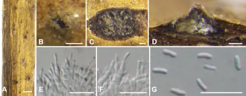
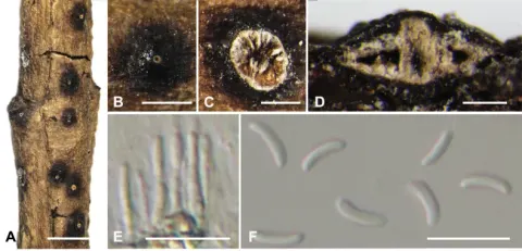
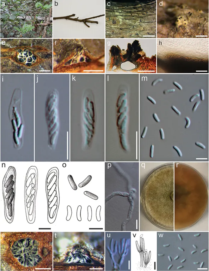
MFLU 17-0708 Associated with Xylocarpus granatum branches. Sexual morph: Stromata immersed in bark. Ascostromata 230–600 × 90–250 µm diameter, semi-immersed in host tissue, scattered, erumpent, multi-loculate, with ostiolar neck. Ostiole 160–200 µm diameter, numerous, dark brown to black, at the same level as the disc, occasionally area surrounded with white hyphae. Peridium comprising several layers of cells of textura angularis, with innermost layer thick, pale brown, outer layer dark brown to black. Hamathecium comprising long cylindrical, cellular, anastomosed paraphyses. Asci (22–)24–28.8 × 3.6–4.8(–5.1) μm (x ‒ = 26 × 4 μm, n = 15), 6–8-spored, unitunicate, clavate to elongate obovoid, with a refractive, J-, apical ring. Ascospores (5.5–)6–6.5 × 1.7–1.8(–2) μm (x ‒ = 5.7 × 1.8 μm, n = 20), biseriate, elongate-allantoid, unicellular hyaline, smooth-walled. Asexual morph: Conidiomata 700–1200 × 400–480 µm diameter, semi-immersed in host tissue, solitary, erumpent, scattered, multi-loculate, with ostiole. Ostioles 200–250 µm long, with 1–2 ostiolar necks. Peridium comprising several layers of cells of textura angularis, with innermost layer brown, outer layer dark brown to black. Conidiophores unbranched or occasionally branched at the bases, formed from the innermost layer of pycnidial wall, with conidiogenous cells. Conidiogenous cells (6.3–)7.9–10 × 0.9–1.4(–1.6) μm (x ‒ = 8.5× 1.4 μm, n = 15), blastic, enteroblastic, flask-shaped, phialidic, hyaline and smooth-walled. Conidia (2.4–)3–3.1 × 0.8–1(–1.2) µm (x ‒ = 3 × 1 µm, n = 30), unicellular, subcylindrical, hyaline, smooth-walled.
Habitat dan peran ekologis
The asexual morph of C. xylocarpi, studied here, is most similar to C. rhizophorae from dead roots of Rhizophora mangle L. in Guatemala, in having multi-loculate conidiomata and allantoid, slightly curved, hyaline and 3–6 × 1.1–1.5 μm conidia (Kohlmeyer and Kohlmeyer 1971). However, the phylogenies, generated herein, show that C. xylocarpi is distinct from C. rhizophorae (ATCC 38475), a strain from Rhizophora mangle that was identified by Kohlmeyer, the author of the species (Fig. 2). The two species also differ by 25 substitutions in ITS1+ITS2 and were collected from different hosts. Therefore, the collection in the present study is designated as a new species. Our phylogeny also indicates a close relationship to unpublished sequences from GenBank (Figs 1, 2). Given that no morphological descriptions are available for these, the similarity in the ITS1 and ITS2 sequence between our strain and the sequences from GenBank (HAB16R13, M225, A761, MUCC302) are presented in Table 3. Those strains were collected from different hosts (Table 3) and, together with our strain, show substantial variation in ITS1 and ITS2 (Table 4). More collections are needed to further study morphological and genetic variation in this group.
- Habitat atau konteks temuan
- Informasi belum tersedia pada sumber rilis ini.
- Substrat
- Informasi belum tersedia pada sumber rilis ini.
- Peran atau konteks
- Belum dikategorikan pada rilis ini.
Reproduksi
MFLU 17-0708 Associated with Xylocarpus granatum branches. Sexual morph: Stromata immersed in bark. Ascostromata 230–600 × 90–250 µm diameter, semi-immersed in host tissue, scattered, erumpent, multi-loculate, with ostiolar neck. Ostiole 160–200 µm diameter, numerous, dark brown to black, at the same level as the disc, occasionally area surrounded with white hyphae. Peridium comprising several layers of cells of textura angularis, with innermost layer thick, pale brown, outer layer dark brown to black. Hamathecium comprising long cylindrical, cellular, anastomosed paraphyses. Asci (22–)24–28.8 × 3.6–4.8(–5.1) μm (x ‒ = 26 × 4 μm, n = 15), 6–8-spored, unitunicate, clavate to elongate obovoid, with a refractive, J-, apical ring.
Catatan identitas dan cakupan
- Nama diterima mengikuti Catalogue of Life Base2026-09-11; uraian mempertahankan konsep dan nama sumber aslinya.
- Preserve original treatment localities, hosts and substrate scope. Materials examined describe documented specimens, not the full global range.
- Impor dan terjemahan otomatis belum diperiksa editorial per profil. Struktur reproduktif yang disebutkan tidak membuktikan seluruh siklus hidup diketahui.
- Pengukuran pada uraian mengikuti spesimen, kultur, dan metode sumber; angka tidak diperluas ke semua populasi.
Catatan distribusi dalam narasi mengikuti sumbernya. Dokumentasi spesimen atau foto mengikuti catatan sumber. Bukti wilayah Indonesia dan penilaian konservasi tidak disimpulkan dari gambar.
Melihat dengan konteks
Cytospora xylocarpi (MFLU 17-0708, holotype). a Xylocarpus granatum b Branch of Xylocarpus granatum c Ascostromata on host substrate d Surface of ascomata e Transverse sections through ascostroma to show distribution of locules f, g Longitudinal sections through ascostroma to show distribution of locules h Peridium i – l, n Asci m, o Ascospores p Germinating spore q, r Colonies on MEA (q-from above, r-below) s Transverse sections through conidioma to show distribution of locules t Longitudinal sections through conidioma to show distribution of locules u, v Conidiogenous cells with attached conidia w Mature conidia. Scale bars: c = 2000 µm, d–f = 500 µm, g = 200 µm, h = 20 µm, i, p = 10 µm, j–o, u–w = 5 µm, s, t = 400 µm.
- Jenis
- Pelat ilmiah
- Kredit
- Chada Norphanphoun, Olivier Raspé, Rajesh Jeewon, Ting-Chi Wen, Kevin D. Hyde
- Lisensi
- CC BY 4.0
- Sumber media
- Buka entri sumber
Sumber profil
- [1] Morphological and phylogenetic characterisation of novel Cytospora species associated with mangroves — Cytospora xylocarpi
- [2] Catalogue of Life Base2026-09-11 — Cytospora xylocarpi
- [3] Dokumentasi visual — Cytospora xylocarpi
Rujukan halaman ini
Fungi. Cytospora xylocarpi. Rilis data fungi-2026-10-12-expansion-2. https://fungi-atlas.mgadib.chatgpt.site/jamur/cytospora-xylocarpi/.
Status: impor sumber; pemeriksaan editorial per profil belum selesai. Untuk klaim biologis, gunakan rujukan pada bagian terkait dan periksa konteks sumber aslinya.