Ciri dan struktur
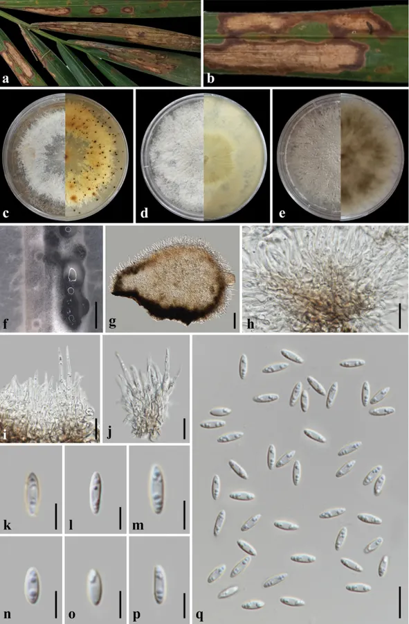
Associated with leaf spots of Nypa fruticans. Leaf spots necrotic, initially round, then expand and elongate to irregularly oval, brown with a darker brown margin. Asexual morph: On alfalfa stem on WA: conidiomata pycnidia 220–460 μm diam., solitary or aggregate, semi-immersed to submerged, black, sub-globose to globose, ostiolate with abundant pale white conidial cirri. Conidiophores 12.5–24.5 × 1.5–3.2 μm (20.2 ± 2.3 × 2.1 ± 0.5 μm, n = 30, l/w: 9.6), hyaline, cylindrical, septate, unbranched, straight, or slightly curved, tapering towards the apex. Conidiogenous cells, 5.5–9 × 1.4–2.2 μm (7.8 ± 0.8 × 1.9 ± 0.2 μm, n = 30, l/w: 4.1), aseptate, hyaline, ellipsoidal to fusoid, and eguttulate. Alpha conidia 6.5–10.5 × 2–3.2 μm (8.2 × 2.5 μm, n = 50, l/w: 3.3), aseptate, hyaline, ellipsoidal to fusoid, multi-guttulate, base acute to acuminate. Beta conidia and gamma conidia not observed. Sexual morph: Not observed. Colonies on PDA media, reaching 52.5 mm, when incubated at room temperatures (28 ± 2 °C) for 7 days, produce three morphotypes: a) white, flat, filamentous, sparse aerial mycelium with circular and irregular margin, and reverse is orange yellow with visible submerged conidiomata; b) white, flat with entire margin, and reverse is yellow; c) gray, flat, floccose to woolly with entire margin and reverse is pale gray with concentric darker gray zones radiating from the center.
Habitat dan peran ekologis
Cosmopolitan. Mangrove species: Annona glabra, Bruguiera spp., Kandelia candel, Lumnitzera spp., Millettia pinnata, Rhizophora spp., Sonneratia spp., Xylocarpus spp.; Arecaceae: Areca catechu, Arenga engleri, Chamaerops humilis, Chrysalidocarpus lutescens, Dypsis lutescens, Livistona chinensis, Phoenix canariensis, Phoenix paludosa, P. dactylifera, Trachycarpus fortunei (Pereira et al. 2023; Aumentado et al. 2024b; Farr and Rossman 2026).
- Habitat atau konteks temuan
- Informasi belum tersedia pada sumber rilis ini.
- Substrat
- Informasi belum tersedia pada sumber rilis ini.
- Peran atau konteks
- Belum dikategorikan pada rilis ini.
Reproduksi
Associated with leaf spots of Nypa fruticans. Leaf spots necrotic, initially round, then expand and elongate to irregularly oval, brown with a darker brown margin. Asexual morph: On alfalfa stem on WA: conidiomata pycnidia 220–460 μm diam., solitary or aggregate, semi-immersed to submerged, black, sub-globose to globose, ostiolate with abundant pale white conidial cirri. Conidiophores 12.5–24.5 × 1.5–3.2 μm (20.2 ± 2.3 × 2.1 ± 0.5 μm, n = 30, l/w: 9.6), hyaline, cylindrical, septate, unbranched, straight, or slightly curved, tapering towards the apex. Conidiogenous cells, 5.5–9 × 1.4–2.2 μm (7.8 ± 0.8 × 1.9 ± 0.2 μm, n = 30, l/w: 4.1), aseptate, hyaline, ellipsoidal to fusoid, and eguttulate.
Catatan identitas dan cakupan
- Nama diterima mengikuti Catalogue of Life Base2026-09-11; uraian mempertahankan konsep dan nama sumber aslinya.
- Preserve original treatment localities, hosts and substrate scope. Materials examined describe documented specimens, not the full global range.
- Impor dan terjemahan otomatis belum diperiksa editorial per profil. Struktur reproduktif yang disebutkan tidak membuktikan seluruh siklus hidup diketahui.
- Pengukuran pada uraian mengikuti spesimen, kultur, dan metode sumber; angka tidak diperluas ke semua populasi.
Catatan distribusi dalam narasi mengikuti sumbernya. Dokumentasi spesimen atau foto mengikuti catatan sumber. Bukti wilayah Indonesia dan penilaian konservasi tidak disimpulkan dari gambar.
Melihat dengan konteks
Diaporthe arecae (MFLUCC 26-0134). a, b. Leaf spot symptoms on Nypa fruticans; c–e. Colony morphotypes on PDA at 1 month post-incubation (obverse, reverse); f. Conidiomata on alfalfa stem in WA; g. Vertical section of conidioma; h–j. Conidial attachment and conidiophores; k–q. Conidia. Scale bars: 1 mm (f); 50 μm (g); 20 μm (h); 10 μm (i–j, q); 5 μm (k–p).
- Jenis
- Pelat ilmiah
- Kredit
- Herbert Dustin R. Aumentado, Ruvishika S. Jayawardena, Carlo Chris Apurillo, Chayanard Phukhamsakda, E. B. Gareth Jones, Eric H. C. McKenzie, Kevin D. Hyde, Long-Feng Yu, Yong Wang
- Lisensi
- CC BY 4.0
- Sumber media
- Buka entri sumber
Dokumentasi tambahan
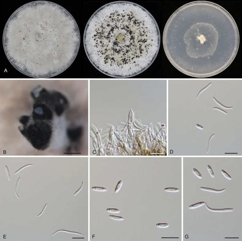
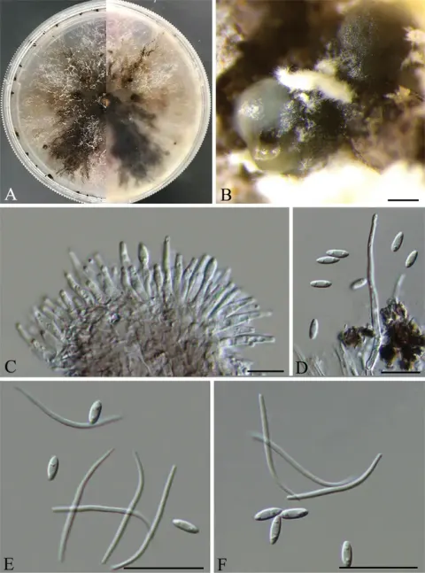
Morphology of Diaporthe arecae (CFCC 55899). A Colonies on PDA, MPA and SNA after 20 d at 25 °C. B Conidiomata. C Conidiogenous cells. D–G Alpha and beta conidia. Scale bars: 400 μm (B); 10 μm (C–G).
Kredit dan penggunaan
Xue Zhong, Hermann Voglmayr, Han Xue, Yong Li, Ning Jiang
Ukuran dan format gambar disesuaikan tanpa pemotongan, retus, atau penambahan skala. Lisensi sumber dipertahankan.
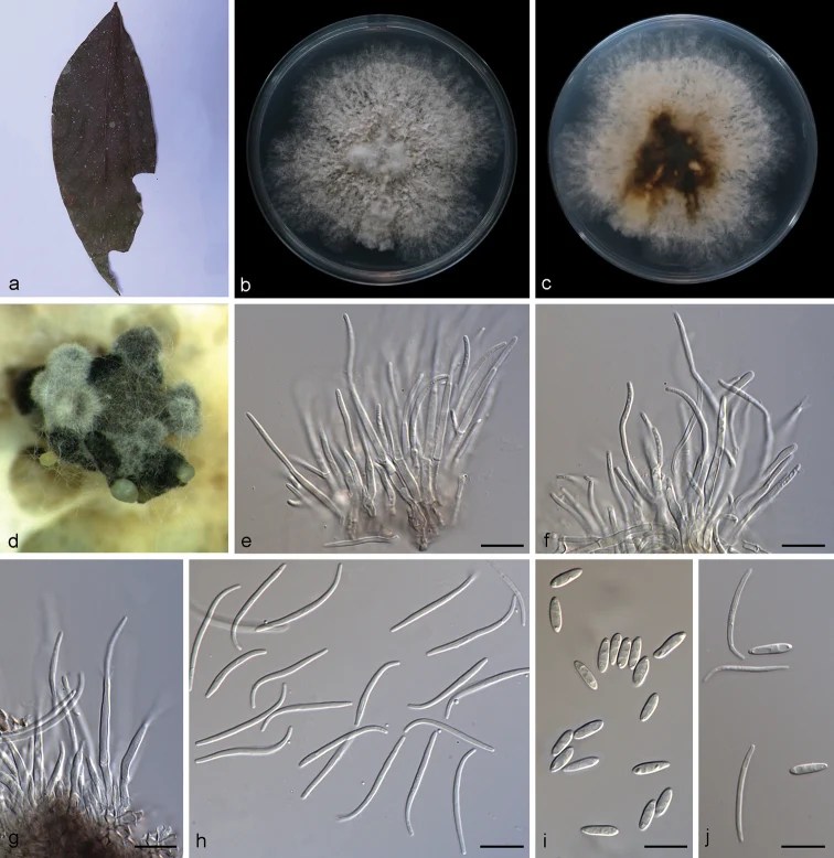
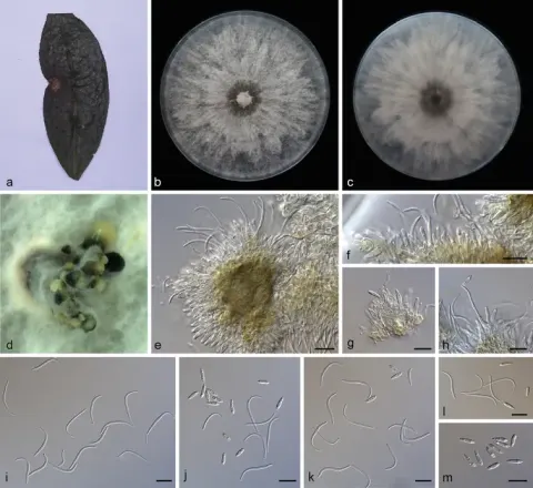
Diaporthe arecae (SAUCC194.18) a infected leaf of Persea americana b, c surface and reverse of a colony after 15 days on PDA d conidiomata e–g conidiophores and conidiogenous cells h beta conidia i alpha conidia j alpha conidia and beta conidia. Scale bars: 10 μm (e–j).
Kredit dan penggunaan
Shengting Huang, Jiwen Xia, Xiuguo Zhang, WenXiu Sun
Ukuran dan format gambar disesuaikan tanpa pemotongan, retus, atau penambahan skala. Lisensi sumber dipertahankan.
Sumber profil
- [1] Putative palm pathogens: Novel species and new host records of leaf disease-associated microfungi (Ascomycota) on Nypa fruticans in Thailand — Diaporthe arecae
- [2] Catalogue of Life Base2026-09-11 — Diaporthe arecae
- [3] Dokumentasi visual — Diaporthe arecae
- [4] Dokumentasi visual — Diaporthe arecae
- [5] Dokumentasi visual — Diaporthe arecae
Rujukan halaman ini
Fungi. Diaporthe arecae. Rilis data fungi-2026-10-12-expansion-2. https://fungi-atlas.mgadib.chatgpt.site/jamur/diaporthe-arecae/.
Status: impor sumber; pemeriksaan editorial per profil belum selesai. Untuk klaim biologis, gunakan rujukan pada bagian terkait dan periksa konteks sumber aslinya.