Ciri dan struktur
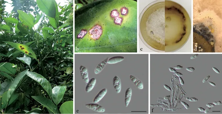
Leaf spots subcircular to irregular, pale brown, with dark brown at margin. Pycnidia solitary, scattered on the leaf surface. Pycnidial conidiomata in culture, globose, erumpent, single or clustered in groups of 3–5 pycnidia, coated with hyphae, cream to yellowish translucent conidial droplets exuded from ostioles. Conidiophores reduced to conidiogenous cells. Conidiogenous cells hyaline, unbranched, septate, straight, slightly tapering towards the apex, 14.5–25 × 2–3 μm. Alpha conidia hyaline, aseptate, rounded at one end, apex at the other end, usually with two large guttulate, (9.5–)10.5–12 × 3.5–4.5 μm. Beta conidia not observed.
Habitat dan peran ekologis
Cunningham (1931b) and Fletcher (1955a) recorded melanose on living leaves and fruits of citrus. Fletcher (1955b) reported that infection was carried over on dead twigs and branches, where the fungus caused dieback, especially of weakened shoots: he recommended a pre-blossom spray of Bordeaux (3:4:50) for its control. This fungus is also responsible for a stem end rot of fruits held in store.
- Habitat atau konteks temuan
- Informasi belum tersedia pada sumber rilis ini.
- Substrat
- Informasi belum tersedia pada sumber rilis ini.
- Peran atau konteks
- Belum dikategorikan pada rilis ini.
Reproduksi
Leaf spots subcircular to irregular, pale brown, with dark brown at margin. Pycnidia solitary, scattered on the leaf surface. Pycnidial conidiomata in culture, globose, erumpent, single or clustered in groups of 3–5 pycnidia, coated with hyphae, cream to yellowish translucent conidial droplets exuded from ostioles. Conidiophores reduced to conidiogenous cells. Conidiogenous cells hyaline, unbranched, septate, straight, slightly tapering towards the apex, 14.5–25 × 2–3 μm. Alpha conidia hyaline, aseptate, rounded at one end, apex at the other end, usually with two large guttulate, (9.5–)10.5–12 × 3.5–4.5 μm. Beta conidia not observed.
Catatan identitas dan cakupan
- Nama diterima mengikuti Catalogue of Life Base2026-09-11; uraian mempertahankan konsep dan nama sumber aslinya.
- Uraian digabungkan dari sumber spesies yang sama; lingkup geografis, bahan, dan kultur setiap sumber dipertahankan. Preserve original treatment localities, hosts and substrate scope. Materials examined describe documented specimens, not the full global range. Historical description as cited by BiotaNZ. Preserve explicit New Zealand/locality, host and specimen qualifiers. Distribution is the cited source scope, not a current global range.
- Impor dan terjemahan otomatis belum diperiksa editorial per profil. Struktur reproduktif yang disebutkan tidak membuktikan seluruh siklus hidup diketahui.
- Pengukuran pada uraian mengikuti spesimen, kultur, dan metode sumber; angka tidak diperluas ke semua populasi.
Catatan distribusi dalam narasi mengikuti sumbernya. Dokumentasi spesimen atau foto mengikuti catatan sumber. Bukti wilayah Indonesia dan penilaian konservasi tidak disimpulkan dari gambar.
Melihat dengan konteks
Diaporthe citri on Citrus sinensis (BJFC -S1658) a, b symptoms on leaves of host plant c culture on PDA (30d) d conidiomata e alpha conidia f conidiophores and alpha conidia. Scale bars: 10 μm (e, f).
- Jenis
- Pelat ilmiah
- Kredit
- Qin Yang, Ning Jiang, Cheng-Ming Tian
- Lisensi
- CC BY 4.0
- Sumber media
- Buka entri sumber
Sumber profil
- [1] New species and records of Diaporthe from Jiangxi Province, China — Diaporthe citri
- [2] Dingley, J.M. 1969: Records of plant diseases in New Zealand. New Zealand Department of Scientific and Industrial Research, Bulletin. 192. — Diaporthe citri
- [3] Catalogue of Life Base2026-09-11 — Diaporthe citri
- [4] Dokumentasi visual — Diaporthe citri
Rujukan halaman ini
Fungi. Diaporthe citri. Rilis data fungi-2026-10-12-expansion-2. https://fungi-atlas.mgadib.chatgpt.site/jamur/diaporthe-citri/.
Status: impor sumber; pemeriksaan editorial per profil belum selesai. Untuk klaim biologis, gunakan rujukan pada bagian terkait dan periksa konteks sumber aslinya.