01 / Morfologi

Ciri dan struktur

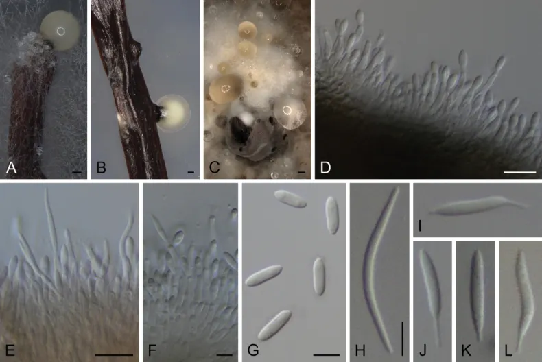
Conidiomata pycnidial in culture on PNA, globose or irregular, dark brown to black, solitary or in groups, embedded, erumpent, 200–460 μm diam, white to cream or yellow conidial drops exuded from ostioles; conidiomatal wall yellowish brown to olivaceous brown or brown, composed of 1–6 layers, textura angularis. Conidiophores cylindrical to subcylindrical, base subhyaline to pale olivaceous, apex hyaline, densely aggregated, smooth-walled, 1–3-septate, 13–42 × 1.5–4 μm. Conidiogenous cells phialidic, cylindrical to subcylindrical, tapering towards apex, hyaline, terminal or lateral, (5.5–)6.5–14 × 1.5–3 μm. Paraphyses not observed. Alpha conidia ellipsoidal to obovoid, or fusoid-ellipsoid, hyaline, apex rounded or subobtuse, base acutate or subtruncate, biguttulate to multiguttulate, aseptate, 5.5–9(–10) × 2–3(–3.5) μm. Beta conidia less frequent, filiform, curved, tapering towards apex, hyaline, not guttulate, aseptate, 11.5–27.5 × 1–2 μm. Gamma conidia less frequent, broadly fusiform, straight to slightly curved, rarely sinuose, apex acutate or filiform, base filiform, hyaline, multiguttulate, aseptate, 10–18.5(–21) × 1.5–2.5 μm.

Sebagian uraian ditampilkan dalam bahasa sumber; terjemahannya belum tersedia atau belum lolos pemeriksaan.

Rujukan: [1]

Pengukuran mengikuti objek, spesimen atau kultur, dan metode yang disebutkan sumber. Baca uraian asli untuk konteks lengkap.

02 / Kehidupan

Habitat dan peran ekologis

Colonies on PDA reaching 63–72 mm or covering the surface of the Petri dish in 2 weeks, white with a grayed yellow (160C) ring and transparent margins, lobate, cottony to slightly feathery, flat to raised in some zones or fully raised, lobate, margins filamentous to fimbriate; reverse grayed yellow (160B–D). Colonies on MEA covering the surface of the Petri dish in 2 weeks, grayed yellow (161A) with a white ring and white to transparent margins, cottony to slightly feathery, flat to raised in some zones or fully raised, margins filamentous to fimbriate; reverse grayed yellow (162A–C) with transparent margins and sometimes with gray brown (199A) center. Colonies on OA covering the surface of the Petri dish in 2 weeks, white to grayed white (156A) with grayed yellow (161A–B) margins or grayed yellow (161C) with brown (200A) dots and white center and margins, cottony to slightly feathery, raised, margins filamentous to fimbriate; reverse grayed green (197B) to/or gray brown (199C–D). Diaporthe isoberliniae was described based on a specimen isolated from Zambia on Isoberlinia angolensis (Fabaceae) (Crous et al. 2014b). To the best of our knowledge, this species had not been recollected since then.

Sebagian uraian ditampilkan dalam bahasa sumber; terjemahannya belum tersedia atau belum lolos pemeriksaan.

Rujukan: [1]

Habitat atau konteks temuan
kultur yang didokumentasikan
Substrat
Informasi belum tersedia pada sumber rilis ini.
Peran atau konteks
Belum dikategorikan pada rilis ini.
Temukan hubungan serupa
03 / Siklus hidup

Reproduksi

Conidiomata pycnidial in culture on PNA, globose or irregular, dark brown to black, solitary or in groups, embedded, erumpent, 200–460 μm diam, white to cream or yellow conidial drops exuded from ostioles; conidiomatal wall yellowish brown to olivaceous brown or brown, composed of 1–6 layers, textura angularis. Conidiophores cylindrical to subcylindrical, base subhyaline to pale olivaceous, apex hyaline, densely aggregated, smooth-walled, 1–3-septate, 13–42 × 1.5–4 μm. Conidiogenous cells phialidic, cylindrical to subcylindrical, tapering towards apex, hyaline, terminal or lateral, (5.5–)6.5–14 × 1.5–3 μm. Paraphyses not observed. Alpha conidia ellipsoidal to obovoid, or fusoid-ellipsoid, hyaline, apex rounded or subobtuse, base acutate or subtruncate, biguttulate to multiguttulate, aseptate, 5.5–9(–10) × 2–3(–3.5) μm.

Sebagian uraian ditampilkan dalam bahasa sumber; terjemahannya belum tersedia atau belum lolos pemeriksaan.

Rujukan: [1]

04 / Batas informasi

Catatan identitas dan cakupan

  • Nama diterima mengikuti Catalogue of Life Base2026-09-11; uraian mempertahankan konsep dan nama sumber aslinya.
  • Preserve original treatment localities, hosts and substrate scope. Materials examined describe documented specimens, not the full global range.
  • Impor dan terjemahan otomatis belum diperiksa editorial per profil. Struktur reproduktif yang disebutkan tidak membuktikan seluruh siklus hidup diketahui.
  • Pengukuran pada uraian mengikuti spesimen, kultur, dan metode sumber; angka tidak diperluas ke semua populasi.

Catatan distribusi dalam narasi mengikuti sumbernya. Dokumentasi spesimen atau foto mengikuti catatan sumber. Bukti wilayah Indonesia dan penilaian konservasi tidak disimpulkan dari gambar.

05 / Dokumentasi

Melihat dengan konteks

Diaporthe isoberliniae A, B conidioma in PNA C conidiomata in OA D–F conidiophores and conidia G alpha conidia H beta conidia I–L Gamma conidia A, C, D–I STMA 18291 B, J–L STMA 18245. Scale bars: 100 μm (A, B); 500 μm (C), 10 μm (D, E), 5 μm (F–L).

Jenis
Pelat ilmiah
Kredit
Christopher Lambert, Lena Schweizer, Blondelle Matio Kemkuignou, Elodie Gisèle M. Anoumedem, Simeon F. Kouam, Yasmina Marin-Felix
Lisensi
CC BY 4.0
Sumber media
Buka entri sumber

Gambar menunjukkan objek menurut identifikasi sumber, bukan verifikasi independen spesimen oleh Fungi. Tidak ada skala baru yang ditambahkan.

06 / Rujukan

Sumber profil

  1. [1] Four new endophytic species of Diaporthe (Diaporthaceae, Diaporthales) isolated from Cameroon — Diaporthe isoberliniaeBagian sumber: PMC10616871_section_31
    Tanggal penghimpunan sumber: 2026-10-12
    Teks sumber: CC BY 4.0 · Lambert C, Schweizer L, Matio Kemkuignou B, Anoumedem EGM, Kouam SF, Marin-Felix Y.
  2. [2] Catalogue of Life Base2026-09-11 — Diaporthe isoberliniaeBagian sumber: ColDP NameUsage.tsv ID 35CWW; accepted species; source 2073
    Tanggal penghimpunan sumber: 2026-10-12
  3. [3] Dokumentasi visual — Diaporthe isoberliniaeBagian sumber: Individual media record; identification follows the source, not independent determination
    Tanggal penghimpunan sumber: 2026-10-12
    Media sumber: CC BY 4.0 · Christopher Lambert, Lena Schweizer, Blondelle Matio Kemkuignou, Elodie Gisèle M. Anoumedem, Simeon F. Kouam, Yasmina Marin-Felix

Adaptasi teks: CC BY 4.0. Lambert C, Schweizer L, Matio Kemkuignou B, Anoumedem EGM, Kouam SF, Marin-Felix Y. — Four new endophytic species of Diaporthe (Diaporthaceae, Diaporthales) isolated from Cameroon — Diaporthe isoberliniae

Uraian asli dan riwayat sumber

Riwayat atau halaman sumber

Ringkasan sumber

Conidiomata pycnidial in culture on PNA, globose or irregular, dark brown to black, solitary or in groups, embedded, erumpent, 200–460 μm diam, white to cream or yellow conidial drops exuded from ostioles; conidiomatal wall yellowish brown to olivaceous brown or brown, composed of 1–6 layers, textura angularis. Conidiophores cylindrical to subcylindrical, base subhyaline to pale olivaceous, apex hyaline, densely aggregated, smooth-walled, 1–3-septate, 13–42 × 1.5–4 μm. Conidiogenous cells phialidic, cylindrical to subcylindrical, tapering towards apex, hyaline, terminal or lateral, (5.5–)6.5–14 × 1.5–3 μm.

Revisi sumber

Morfologi sumber

Conidiomata pycnidial in culture on PNA, globose or irregular, dark brown to black, solitary or in groups, embedded, erumpent, 200–460 μm diam, white to cream or yellow conidial drops exuded from ostioles; conidiomatal wall yellowish brown to olivaceous brown or brown, composed of 1–6 layers, textura angularis. Conidiophores cylindrical to subcylindrical, base subhyaline to pale olivaceous, apex hyaline, densely aggregated, smooth-walled, 1–3-septate, 13–42 × 1.5–4 μm. Conidiogenous cells phialidic, cylindrical to subcylindrical, tapering towards apex, hyaline, terminal or lateral, (5.5–)6.5–14 × 1.5–3 μm. Paraphyses not observed. Alpha conidia ellipsoidal to obovoid, or fusoid-ellipsoid, hyaline, apex rounded or subobtuse, base acutate or subtruncate, biguttulate to multiguttulate, aseptate, 5.5–9(–10) × 2–3(–3.5) μm. Beta conidia less frequent, filiform, curved, tapering towards apex, hyaline, not guttulate, aseptate, 11.5–27.5 × 1–2 μm. Gamma conidia less frequent, broadly fusiform, straight to slightly curved, rarely sinuose, apex acutate or filiform, base filiform, hyaline, multiguttulate, aseptate, 10–18.5(–21) × 1.5–2.5 μm.

Revisi sumber

Ekologi sumber

Colonies on PDA reaching 63–72 mm or covering the surface of the Petri dish in 2 weeks, white with a grayed yellow (160C) ring and transparent margins, lobate, cottony to slightly feathery, flat to raised in some zones or fully raised, lobate, margins filamentous to fimbriate; reverse grayed yellow (160B–D). Colonies on MEA covering the surface of the Petri dish in 2 weeks, grayed yellow (161A) with a white ring and white to transparent margins, cottony to slightly feathery, flat to raised in some zones or fully raised, margins filamentous to fimbriate; reverse grayed yellow (162A–C) with transparent margins and sometimes with gray brown (199A) center. Colonies on OA covering the surface of the Petri dish in 2 weeks, white to grayed white (156A) with grayed yellow (161A–B) margins or grayed yellow (161C) with brown (200A) dots and white center and margins, cottony to slightly feathery, raised, margins filamentous to fimbriate; reverse grayed green (197B) to/or gray brown (199C–D). Diaporthe isoberliniae was described based on a specimen isolated from Zambia on Isoberlinia angolensis (Fabaceae) (Crous et al. 2014b). To the best of our knowledge, this species had not been recollected since then.

Revisi sumber

Struktur reproduksi sumber

Conidiomata pycnidial in culture on PNA, globose or irregular, dark brown to black, solitary or in groups, embedded, erumpent, 200–460 μm diam, white to cream or yellow conidial drops exuded from ostioles; conidiomatal wall yellowish brown to olivaceous brown or brown, composed of 1–6 layers, textura angularis. Conidiophores cylindrical to subcylindrical, base subhyaline to pale olivaceous, apex hyaline, densely aggregated, smooth-walled, 1–3-septate, 13–42 × 1.5–4 μm. Conidiogenous cells phialidic, cylindrical to subcylindrical, tapering towards apex, hyaline, terminal or lateral, (5.5–)6.5–14 × 1.5–3 μm. Paraphyses not observed. Alpha conidia ellipsoidal to obovoid, or fusoid-ellipsoid, hyaline, apex rounded or subobtuse, base acutate or subtruncate, biguttulate to multiguttulate, aseptate, 5.5–9(–10) × 2–3(–3.5) μm.

Revisi sumber

Rujukan halaman ini

Fungi. Diaporthe isoberliniae. Rilis data fungi-2026-10-12-expansion-2. https://fungi-atlas.mgadib.chatgpt.site/jamur/diaporthe-isoberliniae/.

Status: impor sumber; pemeriksaan editorial per profil belum selesai. Untuk klaim biologis, gunakan rujukan pada bagian terkait dan periksa konteks sumber aslinya.