Ciri dan struktur
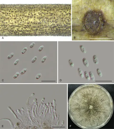
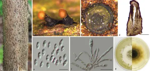
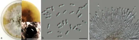
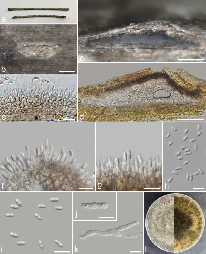
Saprobic on decaying Bambusa. Sexual morph: Undetermined. Asexual morph: Conidiomata 91–148 × 311–974 μm (x̄ = 120 × 583 μm, n = 20), pycnidial, scattered or aggregated, embedded in host surface, slightly erumpent through host surface, 1–3-locular conidioma, nearly flat, elongated, discoid, or variable in shape, black, consisting of hyaline, thin-walled cells of textura angularis, outer layer thick walled. Conidiophores reduced to conidiogenous cells. Conidiogenous cells 4–16 × 1.2–2.6 μm (x̄ = 7.5 × 1.8 μm, n = 40), cylindrical, unbranched, aseptate, smooth, straight or slightly curved, tapering towards the apex, wider at base, hyaline. Alpha conidia 4.1–7.8 × 1.7–3.1 μm (x̄ = 5.8 × 2.5 μm, n = 40), apex bluntly rounded, 1–2-guttulate, mostly bi-guttulate, oval or oblong to ellipsoid, hyaline, smooth, base obtuse to subtruncate. Beta and Gamma conidia not observed. Colonies on PDA, reaching 40 mm diam., after 2 weeks at 25 °C, initially white, turning olivaceous grey after 7–10 days, darker at the centre and marginal area, lacking aerial mycelium; reverse, olivaceous grey bordered by dark margins.
Habitat dan peran ekologis
Actinidia spp. (Actinidiaceae), Alangium kurzii (Cornaceae), Bambusa sp. (Poaceae), Bougainvillea glabra (Nyctaginaceae), Celtis formosana (Ulmaceae), Morinda officinalis (Rutaceae), Tectona grandis (Lamiaceae), Theobroma cacao (Malvaceae), Soil, Vitis vinifera (Vitaceae) (Chang et al. 2005; Crous et al. 2015; Bai et al. 2017; Doilom et al. 2017; Yang et al. 2018; Manawasinghe et al. 2019; Tennakoon et al. 2021; Luo et al. 2022; this study). Australia, China, Korea, Thailand (Chang et al. 2005; Crous et al. 2015; Bai et al. 2017; Doilom et al. 2017; Yang et al. 2018; Manawasinghe et al. 2019; Tennakoon et al. 2021; Luo et al. 2022; this study).
- Habitat atau konteks temuan
- Informasi belum tersedia pada sumber rilis ini.
- Substrat
- Informasi belum tersedia pada sumber rilis ini.
- Peran atau konteks
- Belum dikategorikan pada rilis ini.
Reproduksi
Saprobic on decaying Bambusa. Sexual morph: Undetermined. Asexual morph: Conidiomata 91–148 × 311–974 μm (x̄ = 120 × 583 μm, n = 20), pycnidial, scattered or aggregated, embedded in host surface, slightly erumpent through host surface, 1–3-locular conidioma, nearly flat, elongated, discoid, or variable in shape, black, consisting of hyaline, thin-walled cells of textura angularis, outer layer thick walled. Conidiophores reduced to conidiogenous cells. Conidiogenous cells 4–16 × 1.2–2.6 μm (x̄ = 7.5 × 1.8 μm, n = 40), cylindrical, unbranched, aseptate, smooth, straight or slightly curved, tapering towards the apex, wider at base, hyaline. Alpha conidia 4.1–7.8 × 1.7–3.1 μm (x̄ = 5.8 × 2.5 μm, n = 40), apex bluntly rounded, 1–2-guttulate, mostly bi-guttulate, oval or oblong to ellipsoid, hyaline, smooth, base obtuse to subtruncate.
Catatan identitas dan cakupan
- Nama diterima mengikuti Catalogue of Life Base2026-09-11; uraian mempertahankan konsep dan nama sumber aslinya.
- Preserve original treatment localities, hosts and substrate scope. Materials examined describe documented specimens, not the full global range.
- Impor dan terjemahan otomatis belum diperiksa editorial per profil. Struktur reproduktif yang disebutkan tidak membuktikan seluruh siklus hidup diketahui.
- Pengukuran pada uraian mengikuti spesimen, kultur, dan metode sumber; angka tidak diperluas ke semua populasi.
Catatan distribusi dalam narasi mengikuti sumbernya. Dokumentasi spesimen atau foto mengikuti catatan sumber. Bukti wilayah Indonesia dan penilaian konservasi tidak disimpulkan dari gambar.
Melihat dengan konteks
Diaporthe tulliensis (MFLU 23–0475) a, b conidiomata on host c longitudinal section through conidioma d, e transverse section of conidioma f, g conidiogenous cells giving rise to conidia h, i alpha conidia j, k germinated conidia l front and reverse view of the colony on PDA. Scale bars: 200 µm (b–d); 20 µm (e); 10 µm (f–k).
- Jenis
- Pelat ilmiah
- Kredit
- Chayanard Phukhamsakda, Kevin D. Hyde, Milan C. Samarakoon, Johnny Louangphan, Kedsara Navasit, Fatimah Al-Otibi, Chitrabhanu S. Bhunjun
- Lisensi
- CC BY 4.0
- Sumber media
- Buka entri sumber
Sumber profil
- [1] Unveiling Sordariomycetes taxa associated with woody litter in Northern Thailand — Diaporthe tulliensis
- [2] Catalogue of Life Base2026-09-11 — Diaporthe tulliensis
- [3] Dokumentasi visual — Diaporthe tulliensis
Rujukan halaman ini
Fungi. Diaporthe tulliensis. Rilis data fungi-2026-10-12-expansion-2. https://fungi-atlas.mgadib.chatgpt.site/jamur/diaporthe-tulliensis/.
Status: impor sumber; pemeriksaan editorial per profil belum selesai. Untuk klaim biologis, gunakan rujukan pada bagian terkait dan periksa konteks sumber aslinya.