Ciri dan struktur
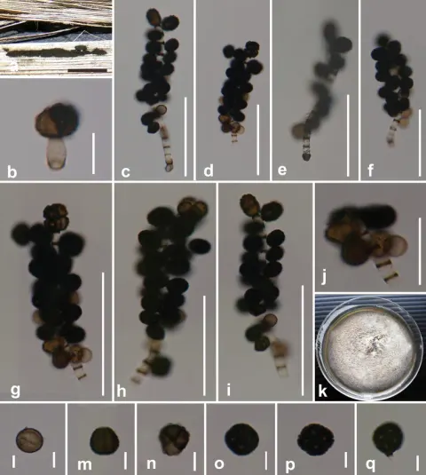
Saprobic on dead leaves of Musa sp. Sexual morph: Undetermined. Asexual morph: Colonies compact or effuse, black, often pulvinate. Mycelium superficial, a close network of branched and anastomosing hyphae. Stromata none. Setae and hyphopodia absent. Conidiophores 30–140 × 1–2 μm (x¯ 81.5 × 1.6 μm, n = 25), basauxic, arising usually singly from subspherical, subhyaline to light brown conidiophore mother cells, 4.5–4.8 × 4.3–4.5 μm (x̄ = 4.6 × 4.4 μm, n = 10), macronematous, mononematous, straight or flexuous, narrow, cylindrical, rough, subhyaline to pale brown, with thick brown or dark brown transverse septa that appear as stripes with distances of 6.3–5.8 μm at apex and 2.3–3 μm at base of the conidiophore. Conidiogenous cells 4.1–4.5 × 4.3–4.7 μm (x̄ = 4.4 × 4.5 μm, n = 10), blastic, integrated, terminal and intercalary, cylindrical, smooth, denticles absent, hyaline. Conidia 7–11.5 × 6.5–9 μm (x̄ = 8.7 × 7.9 μm, n = 40), solitary, dry, acropleurogenous, simple, square, rounded at the corners, 4-celled, spherical or subspherical, often flattened in one plane, pale to dark brown at maturity, verrucose, with light brown to dark brown warts, immature conidia often 1-celled and subhyaline. Terminal conidium with four cells, sometimes absent or fallen before lateral conidia, mature conidia split along one line of the septa, most conidia arranged obliquely downwards on the conidiophore, conidial formation observed as a bunch starting after conidiophore 1–3 septate. Conidia germinating on PDA within 18 hrs. Colonies on PDA reaching a diameter of 50 mm after 14 days at 25 °C, slightly raised, hairy, filamentous, moderately dense, middle light grey, periphery white; reverse white to greyish-white.
Habitat dan peran ekologis
Saprobic on dead leaves of Musa sp. Sexual morph: Undetermined. Asexual morph: Colonies compact or effuse, black, often pulvinate. Mycelium superficial, a close network of branched and anastomosing hyphae. Stromata none. Setae and hyphopodia absent. Conidiophores 30–140 × 1–2 μm (x¯ 81.5 × 1.6 μm, n = 25), basauxic, arising usually singly from subspherical, subhyaline to light brown conidiophore mother cells, 4.5–4.8 × 4.3–4.5 μm (x̄ = 4.6 × 4.4 μm, n = 10), macronematous, mononematous, straight or flexuous, narrow, cylindrical, rough, subhyaline to pale brown, with thick brown or dark brown transverse septa that appear as stripes with distances of 6.3–5.8 μm at apex and 2.3–3 μm at base of the conidiophore. Conidiogenous cells 4.1–4.5 × 4.3–4.7 μm (x̄ = 4.4 × 4.5 μm, n = 10), blastic, integrated, terminal and intercalary, cylindrical, smooth, denticles absent, hyaline. Conidia 7–11.5 × 6.5–9 μm (x̄ = 8.7 × 7.9 μm, n = 40), solitary, dry, acropleurogenous, simple, square, rounded at the corners, 4-celled, spherical or subspherical, often flattened in one plane, pale to dark brown at maturity, verrucose, with light brown to dark brown warts, immature conidia often 1-celled and subhyaline.
- Habitat atau konteks temuan
- Informasi belum tersedia pada sumber rilis ini.
- Substrat
- Informasi belum tersedia pada sumber rilis ini.
- Peran atau konteks
- Pengurai
Reproduksi
Saprobic on dead leaves of Musa sp. Sexual morph: Undetermined. Asexual morph: Colonies compact or effuse, black, often pulvinate. Mycelium superficial, a close network of branched and anastomosing hyphae. Stromata none. Setae and hyphopodia absent. Conidiophores 30–140 × 1–2 μm (x¯ 81.5 × 1.6 μm, n = 25), basauxic, arising usually singly from subspherical, subhyaline to light brown conidiophore mother cells, 4.5–4.8 × 4.3–4.5 μm (x̄ = 4.6 × 4.4 μm, n = 10), macronematous, mononematous, straight or flexuous, narrow, cylindrical, rough, subhyaline to pale brown, with thick brown or dark brown transverse septa that appear as stripes with distances of 6.3–5.8 μm at apex and 2.3–3 μm at base of the conidiophore.
Catatan identitas dan cakupan
- Nama diterima mengikuti Catalogue of Life Base2026-09-11; uraian mempertahankan konsep dan nama sumber aslinya.
- Preserve original treatment localities, hosts and substrate scope. Materials examined describe documented specimens, not the full global range.
- Impor dan terjemahan otomatis belum diperiksa editorial per profil. Struktur reproduktif yang disebutkan tidak membuktikan seluruh siklus hidup diketahui.
- Pengukuran pada uraian mengikuti spesimen, kultur, dan metode sumber; angka tidak diperluas ke semua populasi.
Catatan distribusi dalam narasi mengikuti sumbernya. Dokumentasi spesimen atau foto mengikuti catatan sumber. Bukti wilayah Indonesia dan penilaian konservasi tidak disimpulkan dari gambar.
Melihat dengan konteks
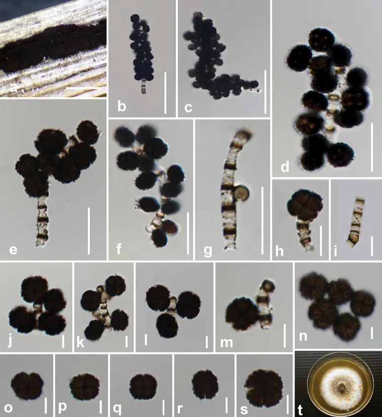
Dictyoarthrinium musae (MFLU 20-0437, holotype) a conidia on the host b conidiophore and conidia with conidiophore mother cell c–f conidia with conidiophores on stalk g developmental stage of an immature lateral conidium h four-celled terminal conidium i conidiophore j conidiophores and conidia with terminal conidium k, l conidiophores without terminal conidium m attachment of a mature lateral conidium n–q warted four-celled mature conidia r, s mature conidia that split at septa t colony on PDA after 21 days. Scale bars: 500 μm (a); 50 μm (b, c); 20 μm (d–g, i); 10 μm (h); 5 μm (j–s).
- Jenis
- Pelat ilmiah
- Kredit
- Binu Samarakoon, Dhanushka N. Wanasinghe, Samantha C. Karunarathna, Milan C. Samarakoon, Rungtiwa Phookamsak, Eric HC Mckenzie, Putarak Chomnunti, Kevin D. Hyde, Saisamorn Lumyong
- Lisensi
- CC BY 4.0
- Sumber media
- Buka entri sumber
Sumber profil
- [1] Multi-gene phylogenetic evidence suggests Dictyoarthrinium belongs in Didymosphaeriaceae (Pleosporales, Dothideomycetes) and Dictyoarthrinium musae sp. nov. on Musa from Thailand — Dictyoarthrinium musae
- [2] Catalogue of Life Base2026-09-11 — Dictyoarthrinium musae
- [3] Dokumentasi visual — Dictyoarthrinium musae
Rujukan halaman ini
Fungi. Dictyoarthrinium musae. Rilis data fungi-2026-10-12-expansion-2. https://fungi-atlas.mgadib.chatgpt.site/jamur/dictyoarthrinium-musae/.
Status: impor sumber; pemeriksaan editorial per profil belum selesai. Untuk klaim biologis, gunakan rujukan pada bagian terkait dan periksa konteks sumber aslinya.