Ciri dan struktur
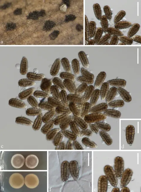
Saprobic on decaying branches in terrestrial habitats. Asexual morph: Hyphomycetous. Colonies on natural substrates scattered, sporodochial, brown to dark brown. Mycelium immersed, composed of branched, septate, pale brown, smooth-walled hyphae. Conidiophores micronematous, mononematous, septate, cylindrical, hyaline to pale brown, smooth-walled, mostly reduced to conidiogenous cells. Conidiogenous cells 4–17 × 3–5 μm (x̄ = 9 × 4.5 μm, n = 20), holoblastic, cylindrical, hyaline to pale brown, smooth-walled. Conidia 42–48 × (15–)18–24 μm (x̄ = 45 × 21 μm, n = 50), solitary, cheiroid, ellipsoid to cylindrical, with a basal connecting cell, pale brown to brown, composed of 5–7 rows of cells, euseptate, unseparated, each row with 10–12 cells, guttulate, slightly constricted at septa, without appendages. Sexual morph: Undetermined. Conidia germinate on PDA within 24 h at room temperature. Germ tubes are produced at the basal region. Colonies on PDA reach 50 mm in diameter after 2 weeks at 20–25 °C; mycelium superficial and circular; surface rough, raised at the center, zonate, grayish white, floccose, and flat; reverse light pink and zonate.
Habitat dan peran ekologis
China and Thailand, on decaying wood from freshwater habitats (Boonmee et al. 2016; Wang et al. 2016; Liu et al. 2023a; Shen et al. 2025) and on decaying branches of Betula albosinensis (Betulaceae) in terrestrial habitats (this study).
- Habitat atau konteks temuan
- Informasi belum tersedia pada sumber rilis ini.
- Substrat
- kayu
- Peran atau konteks
- Belum dikategorikan pada rilis ini.
Reproduksi
Saprobic on decaying branches in terrestrial habitats. Asexual morph: Hyphomycetous. Colonies on natural substrates scattered, sporodochial, brown to dark brown. Mycelium immersed, composed of branched, septate, pale brown, smooth-walled hyphae. Conidiophores micronematous, mononematous, septate, cylindrical, hyaline to pale brown, smooth-walled, mostly reduced to conidiogenous cells. Conidiogenous cells 4–17 × 3–5 μm (x̄ = 9 × 4.5 μm, n = 20), holoblastic, cylindrical, hyaline to pale brown, smooth-walled. Conidia 42–48 × (15–)18–24 μm (x̄ = 45 × 21 μm, n = 50), solitary, cheiroid, ellipsoid to cylindrical, with a basal connecting cell, pale brown to brown, composed of 5–7 rows of cells, euseptate, unseparated, each row with 10–12 cells, guttulate, slightly constricted at septa, without appendages.
Catatan identitas dan cakupan
- Nama diterima mengikuti Catalogue of Life Base2026-09-11; uraian mempertahankan konsep dan nama sumber aslinya.
- Preserve original treatment localities, hosts and substrate scope. Materials examined describe documented specimens, not the full global range.
- Impor dan terjemahan otomatis belum diperiksa editorial per profil. Struktur reproduktif yang disebutkan tidak membuktikan seluruh siklus hidup diketahui.
- Pengukuran pada uraian mengikuti spesimen, kultur, dan metode sumber; angka tidak diperluas ke semua populasi.
Catatan distribusi dalam narasi mengikuti sumbernya. Dokumentasi spesimen atau foto mengikuti catatan sumber. Bukti wilayah Indonesia dan penilaian konservasi tidak disimpulkan dari gambar.
Melihat dengan konteks
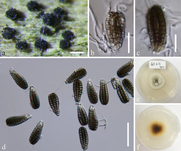
Dictyocheirospora rotunda (MFLU 18-1041). a Colonies on submerged wood b, c Germinated conidia d Conidia e, f Culture, e from above, f from reverse. Scale bars: a = 200 μm, b, c = 20 μm, d = 50 μm.
- Jenis
- Pelat ilmiah
- Kredit
- Jing Yang, Jian-Kui Liu, Kevin D. Hyde, E.B. Gareth Jones, Zuo-Yi Liu
- Lisensi
- CC BY 4.0
- Sumber media
- Buka entri sumber
Dokumentasi tambahan
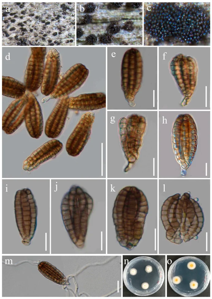
Dictyocheirospora rotunda (HUEST 23.0219, new host record). (a – c) Colonies on a woody substrate. (d) Squash mount of a sporodochium. (e – h) Conidia with partial conidiophores. (i – l) Conidia. (m) Germinated conidium. (n, o) Colonies on PDA, from above (n), from below (o). Scale bars: (d) = 50 μm, (e – l) = 30 μm, (m) = 20 μm.
Kredit dan penggunaan
© 2024 by the authors.
Ukuran dan format gambar disesuaikan tanpa pemotongan, retus, atau penambahan skala. Lisensi sumber dipertahankan.
Sumber profil
- [1] Morphology and multigene phylogeny reveal five new species and two new records of saprobic microfungi from Yunnan, China — Dictyocheirospora rotunda
- [2] Catalogue of Life Base2026-09-11 — Dictyocheirospora rotunda
- [3] Dokumentasi visual — Dictyocheirospora rotunda
- [4] Dokumentasi visual — Dictyocheirospora rotunda
Rujukan halaman ini
Fungi. Dictyocheirospora rotunda. Rilis data fungi-2026-10-12-expansion-2. https://fungi-atlas.mgadib.chatgpt.site/jamur/dictyocheirospora-rotunda/.
Status: impor sumber; pemeriksaan editorial per profil belum selesai. Untuk klaim biologis, gunakan rujukan pada bagian terkait dan periksa konteks sumber aslinya.