Ciri dan struktur
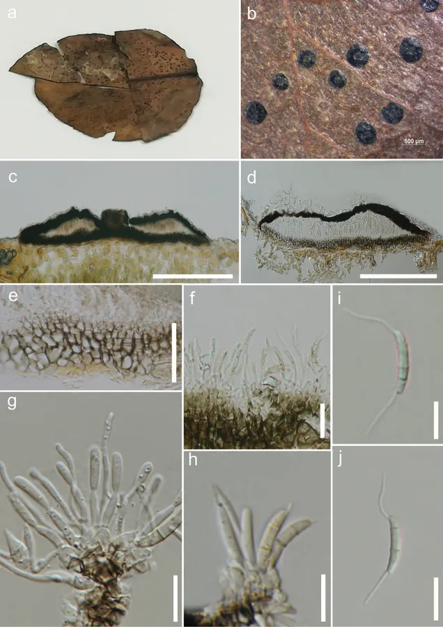
Saprobic on leaves of Pyrus sp. Sexual morph: Undetermined. Asexual morph: Conidiomata 45–70 µm high, 410–800 µm diam., stromatic, scattered to gregarious, superficial, rounded to unevenly outlined with complete margins, applanate, unilocular to bilocular, rugose, not glabrous, dull black, ostiolate. Ostiole 50–90 µm diam., circular to oval, opening to the exterior, central. Conidiomatal wall 10–20 µm thick at the base, dark brown in the outermost layer, comprising thick-walled cells of textura angularis, gradually becoming pale towards the inner layer; 10–20 µm thick near the apex, dark brown to black, made up of thick-walled cells of textura epidermoidea; interlocular wall composed of dark brown thick-walled cells of textura prismatica, becoming thin-walled and paler towards the outer layers. Conidiophores up to 40 µm high, originating from the innermost layer cells of the basal stroma, unbranched or at times branched, mostly 0–1-septate, rarely 2-septate or reduced to conidiogenous cells, cylindrical, hyaline, smooth. Conidiogenous cells 8–30 × 0.7–1.5 µm (x– = 14.3 × 1.1 µm, n = 15), subcylindrical to elongate-ampuliform, hyaline, smooth-walled, holoblastic. Conidia 12–16 × 1.5–3 µm (x– = 13.8 × 2.3 µm, n = 40) naviculate, to subcylindrical, narrow towards the base, straight or faintly curved, euseptate, mostly 3-septate, occasionally 2-septate, with septa thicker and darker than the periclinal wall, with cells unequal, hyaline to sub-hyaline, smooth-walled, without constriction at septa, bearing appendages on both apical and basal cells; basal cell 3–6 µm (x– = 3.8 µm) long, narrowly obconic, with truncate base bearing a conspicuous dehiscence scar; 2 median cells, together 6–10 µm (x– = 7.4 µm) long [second cell 4–6 µm (x– = 5.0 µm) long, close to apical cell, almost twice the size of the third cell 2–4 µm (x– = 3.0 µm) long, close to basal cell]; apical cell 3–5 µm (x– = 3.6 µm) long, subconical with acute apex, hyaline at apex and sub-hyaline below; appendages tubular, faintly broad at the base, unbranched, flexuous; appendage on apical cell 5–17 µm (x– = 10.1 µm) long, single, polar; appendage on basal cell 4–17 µm (x– = 9.4 µm) long, single, inserted slightly above conidium base.
Habitat dan peran ekologis
Saprobic on leaves of Pyrus sp. Sexual morph: Undetermined. Asexual morph: Conidiomata 45–70 µm high, 410–800 µm diam., stromatic, scattered to gregarious, superficial, rounded to unevenly outlined with complete margins, applanate, unilocular to bilocular, rugose, not glabrous, dull black, ostiolate. Ostiole 50–90 µm diam., circular to oval, opening to the exterior, central. Conidiomatal wall 10–20 µm thick at the base, dark brown in the outermost layer, comprising thick-walled cells of textura angularis, gradually becoming pale towards the inner layer; 10–20 µm thick near the apex, dark brown to black, made up of thick-walled cells of textura epidermoidea; interlocular wall composed of dark brown thick-walled cells of textura prismatica, becoming thin-walled and paler towards the outer layers. Conidiophores up to 40 µm high, originating from the innermost layer cells of the basal stroma, unbranched or at times branched, mostly 0–1-septate, rarely 2-septate or reduced to conidiogenous cells, cylindrical, hyaline, smooth. Conidiogenous cells 8–30 × 0.7–1.5 µm (x– = 14.3 × 1.1 µm, n = 15), subcylindrical to elongate-ampuliform, hyaline, smooth-walled, holoblastic. Conidia 12–16 × 1.5–3 µm (x– = 13.8 × 2.3 µm, n = 40) naviculate, to subcylindrical, narrow towards the base, straight or faintly curved, euseptate, mostly 3-septate, occasionally 2-septate, with septa thicker and darker than the periclinal wall, with cells unequal, hyaline to sub-hyaline, smooth-walled, without constriction at septa, bearing appendages on both apical and basal cells; basal cell 3–6 µm (x– = 3.8 µm) long, narrowly obconic, with truncate base bearing a conspicuous dehiscence scar; 2 median cells, together 6–10 µm (x– = 7.4 µm) long [second cell 4–6 µm (x– = 5.0 µm) long, close to apical cell, almost twice the size of the third cell 2–4 µm (x– = 3.0 µm) long, close to basal cell]; apical cell 3–5 µm (x– = 3.6 µm) long, subconical with acute apex, hyaline at apex and sub-hyaline below; appendages tubular, faintly broad at the base, unbranched, flexuous; appendage on apical cell 5–17 µm (x– = 10.1 µm) long, single, polar; appendage on basal cell 4–17 µm (x– = 9.4 µm) long, single, inserted slightly above conidium base.
- Habitat atau konteks temuan
- Informasi belum tersedia pada sumber rilis ini.
- Substrat
- Informasi belum tersedia pada sumber rilis ini.
- Peran atau konteks
- Pengurai
Reproduksi
Saprobic on leaves of Pyrus sp. Sexual morph: Undetermined. Asexual morph: Conidiomata 45–70 µm high, 410–800 µm diam., stromatic, scattered to gregarious, superficial, rounded to unevenly outlined with complete margins, applanate, unilocular to bilocular, rugose, not glabrous, dull black, ostiolate. Ostiole 50–90 µm diam., circular to oval, opening to the exterior, central. Conidiomatal wall 10–20 µm thick at the base, dark brown in the outermost layer, comprising thick-walled cells of textura angularis, gradually becoming pale towards the inner layer; 10–20 µm thick near the apex, dark brown to black, made up of thick-walled cells of textura epidermoidea; interlocular wall composed of dark brown thick-walled cells of textura prismatica, becoming thin-walled and paler towards the outer layers.
Catatan identitas dan cakupan
- Nama diterima mengikuti Catalogue of Life Base2026-09-11; uraian mempertahankan konsep dan nama sumber aslinya.
- Preserve original treatment localities, hosts and substrate scope. Materials examined describe documented specimens, not the full global range.
- Impor dan terjemahan otomatis belum diperiksa editorial per profil. Struktur reproduktif yang disebutkan tidak membuktikan seluruh siklus hidup diketahui.
- Pengukuran pada uraian mengikuti spesimen, kultur, dan metode sumber; angka tidak diperluas ke semua populasi.
Catatan distribusi dalam narasi mengikuti sumbernya. Dokumentasi spesimen atau foto mengikuti catatan sumber. Bukti wilayah Indonesia dan penilaian konservasi tidak disimpulkan dari gambar.
Melihat dengan konteks
Discosia ravennica (MFLU 18-0131, holotype) a Herbarium specimen b Conidiomata on the host c, d Vertical sections of conidiomata e Conidioma wall at the base f–h Conidiogenous cells and developing conidia i, j Conidia. Scale bars: 500 μm (b); 200 μm (c, d); 10 μm (e, g–j); 20 μm (f).
- Jenis
- Pelat ilmiah
- Kredit
- Digvijayini Bundhun, Rajesh Jeewon, Indunil C. Senanayake, Erio Camporesi, Janith V. S. Aluthmuhandiram, Alvin M. C. Tang, Ji-Chuan Kang, Vishwakalyan Bhoyroo, Kevin D. Hyde
- Lisensi
- CC BY 4.0
- Sumber media
- Buka entri sumber
Sumber profil
- [1] Morpho-molecular characterization of Discosia ravennica sp. nov. and a new host record for Sporocadus rosigena — Discosia ravennica
- [2] Catalogue of Life Base2026-09-11 — Discosia ravennica
- [3] Dokumentasi visual — Discosia ravennica
Rujukan halaman ini
Fungi. Discosia ravennica. Rilis data fungi-2026-10-12-expansion-2. https://fungi-atlas.mgadib.chatgpt.site/jamur/discosia-ravennica/.
Status: impor sumber; pemeriksaan editorial per profil belum selesai. Untuk klaim biologis, gunakan rujukan pada bagian terkait dan periksa konteks sumber aslinya.