01 / Morfologi

Ciri dan struktur

Saprobic on submerged decaying wood in a freshwater stream. Anamorph: Colonies on wood effuse, hairy, dark brown, glistening, solitary or in small group. Mycelium immersed, composed of septate, pale brown to brown hyphae, smooth-walled. Conidiophores (42–)66–103(–115) × 5–6 µm (x̄ = 84 × 6 µm, n = 20), macronematous, mononematous, solitary or in groups, erect, straight or slightly flexuous, cylindrical, unbranched, 4–6-septate, brown, rounded at the apex, slightly enlarged at the base, smooth-walled. Conidiogenous cells (10–)15–22(–25) × 5–6 µm (x̄ = 19 × 5 µm, n = 20), monoblastic, terminal, determinate, subcylindrical, brown, smooth-walled. Conidia (55–)69–126(–168) × 10–12 µm (x̄ = 98 × 11 µm, n = 25), acrogenous, solitary, obclavate, olivaceous to pale or dark brown, truncate at base, tapering towards the apex, straight or slightly curved, constricted at the septa, 7–18-distoseptate, guttulate, thick and smooth-walled. Teleomorph: Undetermined. Conidia germinating on PDA within 12 hrs and germ tubes produced from apex and septa of conidium. Colonies growing on PDA reaching 4–5 cm in 20 days at 26 °C in the dark, with dense, velvety, middle papillae, pale to dark brown mycelium from above; dark brown from below.

Sebagian uraian ditampilkan dalam bahasa sumber; terjemahannya belum tersedia atau belum lolos pemeriksaan.

Rujukan: [1]

Pengukuran mengikuti objek, spesimen atau kultur, dan metode yang disebutkan sumber. Baca uraian asli untuk konteks lengkap.

02 / Kehidupan

Habitat dan peran ekologis

Saprobic on submerged decaying wood in a freshwater stream. Anamorph: Colonies on wood effuse, hairy, dark brown, glistening, solitary or in small group. Mycelium immersed, composed of septate, pale brown to brown hyphae, smooth-walled. Conidiophores (42–)66–103(–115) × 5–6 µm (x̄ = 84 × 6 µm, n = 20), macronematous, mononematous, solitary or in groups, erect, straight or slightly flexuous, cylindrical, unbranched, 4–6-septate, brown, rounded at the apex, slightly enlarged at the base, smooth-walled. Conidiogenous cells (10–)15–22(–25) × 5–6 µm (x̄ = 19 × 5 µm, n = 20), monoblastic, terminal, determinate, subcylindrical, brown, smooth-walled. Conidia (55–)69–126(–168) × 10–12 µm (x̄ = 98 × 11 µm, n = 25), acrogenous, solitary, obclavate, olivaceous to pale or dark brown, truncate at base, tapering towards the apex, straight or slightly curved, constricted at the septa, 7–18-distoseptate, guttulate, thick and smooth-walled. Teleomorph: Undetermined. Conidia germinating on PDA within 12 hrs and germ tubes produced from apex and septa of conidium. Colonies growing on PDA reaching 4–5 cm in 20 days at 26 °C in the dark, with dense, velvety, middle papillae, pale to dark brown mycelium from above; dark brown from below.

Sebagian uraian ditampilkan dalam bahasa sumber; terjemahannya belum tersedia atau belum lolos pemeriksaan.

Rujukan: [1]

Habitat atau konteks temuan
kultur yang didokumentasikan
Substrat
kayu
Peran atau konteks
Pengurai
Temukan hubungan serupa
03 / Siklus hidup

Reproduksi

Saprobic on submerged decaying wood in a freshwater stream. Anamorph: Colonies on wood effuse, hairy, dark brown, glistening, solitary or in small group. Mycelium immersed, composed of septate, pale brown to brown hyphae, smooth-walled. Conidiophores (42–)66–103(–115) × 5–6 µm (x̄ = 84 × 6 µm, n = 20), macronematous, mononematous, solitary or in groups, erect, straight or slightly flexuous, cylindrical, unbranched, 4–6-septate, brown, rounded at the apex, slightly enlarged at the base, smooth-walled. Conidiogenous cells (10–)15–22(–25) × 5–6 µm (x̄ = 19 × 5 µm, n = 20), monoblastic, terminal, determinate, subcylindrical, brown, smooth-walled. Conidia (55–)69–126(–168) × 10–12 µm (x̄ = 98 × 11 µm, n = 25), acrogenous, solitary, obclavate, olivaceous to pale or dark brown, truncate at base, tapering towards the apex, straight or slightly curved, constricted at the septa, 7–18-distoseptate, guttulate, thick and smooth-walled.

Sebagian uraian ditampilkan dalam bahasa sumber; terjemahannya belum tersedia atau belum lolos pemeriksaan.

Rujukan: [1]

04 / Batas informasi

Catatan identitas dan cakupan

  • Nama diterima mengikuti Catalogue of Life Base2026-09-11; uraian mempertahankan konsep dan nama sumber aslinya.
  • Preserve original treatment localities, hosts and substrate scope. Materials examined describe documented specimens, not the full global range.
  • Impor dan terjemahan otomatis belum diperiksa editorial per profil. Struktur reproduktif yang disebutkan tidak membuktikan seluruh siklus hidup diketahui.
  • Pengukuran pada uraian mengikuti spesimen, kultur, dan metode sumber; angka tidak diperluas ke semua populasi.

Catatan distribusi dalam narasi mengikuti sumbernya. Dokumentasi spesimen atau foto mengikuti catatan sumber. Bukti wilayah Indonesia dan penilaian konservasi tidak disimpulkan dari gambar.

05 / Dokumentasi

Melihat dengan konteks

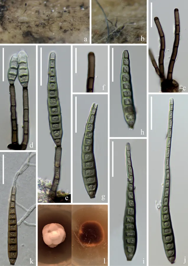
Distoseptispora obpyriformis (HKAS 125823) a, b colonies on woody substrates c conidiophores d, e conidiophores with conidia f conidiogenous cell g–j conidia k germinating conidium l culture on PDA. Scale bars: 30 μm (c–e, g–k); 20 μm (j).

Jenis
Pelat ilmiah
Kredit
Hong-Wei Shen, Dan-Feng Bao, Saranyaphat Boonmee, Yong-Zhong Lu, Xi-Jun Su, Yun-Xia Li, Zong-Long Luo
Lisensi
CC BY 4.0
Sumber media
Buka entri sumber

Gambar menunjukkan objek menurut identifikasi sumber, bukan verifikasi independen spesimen oleh Fungi. Tidak ada skala baru yang ditambahkan.

06 / Rujukan

Sumber profil

  1. [1] Diversity of Distoseptispora (Distoseptisporaceae) taxa on submerged decaying wood from the Red River in Yunnan, China — Distoseptispora obpyriformisBagian sumber: PMC10862349_section_18
    Tanggal penghimpunan sumber: 2026-10-12
    Teks sumber: CC BY 4.0 · Shen HW, Bao DF, Boonmee S, Lu YZ, Su XJ, Li YX, Luo ZL.
  2. [2] Catalogue of Life Base2026-09-11 — Distoseptispora obpyriformisBagian sumber: ColDP NameUsage.tsv ID 36YFQ; accepted species; source 2073
    Tanggal penghimpunan sumber: 2026-10-12
  3. [3] Dokumentasi visual — Distoseptispora obpyriformisBagian sumber: Individual media record; identification follows the source, not independent determination
    Tanggal penghimpunan sumber: 2026-10-12
    Media sumber: CC BY 4.0 · Hong-Wei Shen, Dan-Feng Bao, Saranyaphat Boonmee, Yong-Zhong Lu, Xi-Jun Su, Yun-Xia Li, Zong-Long Luo

Adaptasi teks: CC BY 4.0. Shen HW, Bao DF, Boonmee S, Lu YZ, Su XJ, Li YX, Luo ZL. — Diversity of Distoseptispora (Distoseptisporaceae) taxa on submerged decaying wood from the Red River in Yunnan, China — Distoseptispora obpyriformis

Uraian asli dan riwayat sumber

Riwayat atau halaman sumber

Ringkasan sumber

Saprobic on submerged decaying wood in a freshwater stream. Anamorph: Colonies on wood effuse, hairy, dark brown, glistening, solitary or in small group. Mycelium immersed, composed of septate, pale brown to brown hyphae, smooth-walled. Conidiophores (42–)66–103(–115) × 5–6 µm (x̄ = 84 × 6 µm, n = 20), macronematous, mononematous, solitary or in groups, erect, straight or slightly flexuous, cylindrical, unbranched, 4–6-septate, brown, rounded at the apex, slightly enlarged at the base, smooth-walled.

Revisi sumber

Morfologi sumber

Saprobic on submerged decaying wood in a freshwater stream. Anamorph: Colonies on wood effuse, hairy, dark brown, glistening, solitary or in small group. Mycelium immersed, composed of septate, pale brown to brown hyphae, smooth-walled. Conidiophores (42–)66–103(–115) × 5–6 µm (x̄ = 84 × 6 µm, n = 20), macronematous, mononematous, solitary or in groups, erect, straight or slightly flexuous, cylindrical, unbranched, 4–6-septate, brown, rounded at the apex, slightly enlarged at the base, smooth-walled. Conidiogenous cells (10–)15–22(–25) × 5–6 µm (x̄ = 19 × 5 µm, n = 20), monoblastic, terminal, determinate, subcylindrical, brown, smooth-walled. Conidia (55–)69–126(–168) × 10–12 µm (x̄ = 98 × 11 µm, n = 25), acrogenous, solitary, obclavate, olivaceous to pale or dark brown, truncate at base, tapering towards the apex, straight or slightly curved, constricted at the septa, 7–18-distoseptate, guttulate, thick and smooth-walled. Teleomorph: Undetermined. Conidia germinating on PDA within 12 hrs and germ tubes produced from apex and septa of conidium. Colonies growing on PDA reaching 4–5 cm in 20 days at 26 °C in the dark, with dense, velvety, middle papillae, pale to dark brown mycelium from above; dark brown from below.

Revisi sumber

Ekologi sumber

Saprobic on submerged decaying wood in a freshwater stream. Anamorph: Colonies on wood effuse, hairy, dark brown, glistening, solitary or in small group. Mycelium immersed, composed of septate, pale brown to brown hyphae, smooth-walled. Conidiophores (42–)66–103(–115) × 5–6 µm (x̄ = 84 × 6 µm, n = 20), macronematous, mononematous, solitary or in groups, erect, straight or slightly flexuous, cylindrical, unbranched, 4–6-septate, brown, rounded at the apex, slightly enlarged at the base, smooth-walled. Conidiogenous cells (10–)15–22(–25) × 5–6 µm (x̄ = 19 × 5 µm, n = 20), monoblastic, terminal, determinate, subcylindrical, brown, smooth-walled. Conidia (55–)69–126(–168) × 10–12 µm (x̄ = 98 × 11 µm, n = 25), acrogenous, solitary, obclavate, olivaceous to pale or dark brown, truncate at base, tapering towards the apex, straight or slightly curved, constricted at the septa, 7–18-distoseptate, guttulate, thick and smooth-walled. Teleomorph: Undetermined. Conidia germinating on PDA within 12 hrs and germ tubes produced from apex and septa of conidium. Colonies growing on PDA reaching 4–5 cm in 20 days at 26 °C in the dark, with dense, velvety, middle papillae, pale to dark brown mycelium from above; dark brown from below.

Revisi sumber

Struktur reproduksi sumber

Saprobic on submerged decaying wood in a freshwater stream. Anamorph: Colonies on wood effuse, hairy, dark brown, glistening, solitary or in small group. Mycelium immersed, composed of septate, pale brown to brown hyphae, smooth-walled. Conidiophores (42–)66–103(–115) × 5–6 µm (x̄ = 84 × 6 µm, n = 20), macronematous, mononematous, solitary or in groups, erect, straight or slightly flexuous, cylindrical, unbranched, 4–6-septate, brown, rounded at the apex, slightly enlarged at the base, smooth-walled. Conidiogenous cells (10–)15–22(–25) × 5–6 µm (x̄ = 19 × 5 µm, n = 20), monoblastic, terminal, determinate, subcylindrical, brown, smooth-walled. Conidia (55–)69–126(–168) × 10–12 µm (x̄ = 98 × 11 µm, n = 25), acrogenous, solitary, obclavate, olivaceous to pale or dark brown, truncate at base, tapering towards the apex, straight or slightly curved, constricted at the septa, 7–18-distoseptate, guttulate, thick and smooth-walled.

Revisi sumber

Rujukan halaman ini

Fungi. Distoseptispora obpyriformis. Rilis data fungi-2026-10-12-expansion-2. https://fungi-atlas.mgadib.chatgpt.site/jamur/distoseptispora-obpyriformis/.

Status: impor sumber; pemeriksaan editorial per profil belum selesai. Untuk klaim biologis, gunakan rujukan pada bagian terkait dan periksa konteks sumber aslinya.