Ciri dan struktur
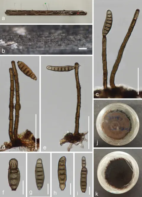
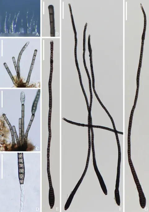
Saprobic on decaying bamboo culms submerged in freshwater habitats. Sexual morph: Undetermined. Asexual morph: Hyphomycetous. Colonies effuse, olivaceous or dark brown, hairy, velvety. Mycelium mostly immersed, consisting of branched, septate, smooth, subhyaline to pale brown hyphae. Conidiophores 100–175 μm × 5.5–10 μm (x‒ = 129×7.1 μm, n = 30), single or in groups of 2 or 3, macronematous, mononematous, erect, straight or slightly flexuous, 4–7-septate, unbranched, olivaceous to dark brown, smooth, cylindrical, rounded at the apex. Conidiogenous cells monoblastic, integrated, terminal, determinate, pale to dark brown, cylindrical. Conidia 39‒67.5(‒77) μm × (7–)9.5–13.5(–16.5) μm (x‒ = 52 × 12 μm, n = 30), acrogenous, solitary, obpyriform or obclavate, thick-walled, tapering towards the rounded apex, slightly curved, truncate at the base, 7–13-distoseptate, guttulate, smooth-walled, olivaceous, dark brown when mature, sometimes with the percurrent proliferation which forms another conidium from the conidial apex. Conidia germinating on PDA within 24 h and germ tubes produced from both ends. Colonies on PDA reaching 12–18 mm diam. at 14 days at 25 °C, in natural light, with fluffy, dense, thin olivaceous mycelium in the centre, becoming sparse and paler at the entire margin; reverse dark brown, pale brown at the smooth margin.
Habitat dan peran ekologis
Saprobic on decaying bamboo culms submerged in freshwater habitats. Sexual morph: Undetermined. Asexual morph: Hyphomycetous. Colonies effuse, olivaceous or dark brown, hairy, velvety. Mycelium mostly immersed, consisting of branched, septate, smooth, subhyaline to pale brown hyphae. Conidiophores 100–175 μm × 5.5–10 μm (x‒ = 129×7.1 μm, n = 30), single or in groups of 2 or 3, macronematous, mononematous, erect, straight or slightly flexuous, 4–7-septate, unbranched, olivaceous to dark brown, smooth, cylindrical, rounded at the apex. Conidiogenous cells monoblastic, integrated, terminal, determinate, pale to dark brown, cylindrical. Conidia 39‒67.5(‒77) μm × (7–)9.5–13.5(–16.5) μm (x‒ = 52 × 12 μm, n = 30), acrogenous, solitary, obpyriform or obclavate, thick-walled, tapering towards the rounded apex, slightly curved, truncate at the base, 7–13-distoseptate, guttulate, smooth-walled, olivaceous, dark brown when mature, sometimes with the percurrent proliferation which forms another conidium from the conidial apex. Conidia germinating on PDA within 24 h and germ tubes produced from both ends. Colonies on PDA reaching 12–18 mm diam. at 14 days at 25 °C, in natural light, with fluffy, dense, thin olivaceous mycelium in the centre, becoming sparse and paler at the entire margin; reverse dark brown, pale brown at the smooth margin.
- Habitat atau konteks temuan
- kultur yang didokumentasikan
- Substrat
- Informasi belum tersedia pada sumber rilis ini.
- Peran atau konteks
- Pengurai
Reproduksi
Saprobic on decaying bamboo culms submerged in freshwater habitats. Sexual morph: Undetermined. Asexual morph: Hyphomycetous. Colonies effuse, olivaceous or dark brown, hairy, velvety. Mycelium mostly immersed, consisting of branched, septate, smooth, subhyaline to pale brown hyphae. Conidiophores 100–175 μm × 5.5–10 μm (x‒ = 129×7.1 μm, n = 30), single or in groups of 2 or 3, macronematous, mononematous, erect, straight or slightly flexuous, 4–7-septate, unbranched, olivaceous to dark brown, smooth, cylindrical, rounded at the apex. Conidiogenous cells monoblastic, integrated, terminal, determinate, pale to dark brown, cylindrical. Conidia 39‒67.5(‒77) μm × (7–)9.5–13.5(–16.5) μm (x‒ = 52 × 12 μm, n = 30), acrogenous, solitary, obpyriform or obclavate, thick-walled, tapering towards the rounded apex, slightly curved, truncate at the base, 7–13-distoseptate, guttulate, smooth-walled, olivaceous, dark brown when mature, sometimes with the percurrent proliferation which forms another conidium from the conidial apex.
Catatan identitas dan cakupan
- Nama diterima mengikuti Catalogue of Life Base2026-09-11; uraian mempertahankan konsep dan nama sumber aslinya.
- Preserve original treatment localities, hosts and substrate scope. Materials examined describe documented specimens, not the full global range.
- Impor dan terjemahan otomatis belum diperiksa editorial per profil. Struktur reproduktif yang disebutkan tidak membuktikan seluruh siklus hidup diketahui.
- Pengukuran pada uraian mengikuti spesimen, kultur, dan metode sumber; angka tidak diperluas ke semua populasi.
Catatan distribusi dalam narasi mengikuti sumbernya. Dokumentasi spesimen atau foto mengikuti catatan sumber. Bukti wilayah Indonesia dan penilaian konservasi tidak disimpulkan dari gambar.
Melihat dengan konteks
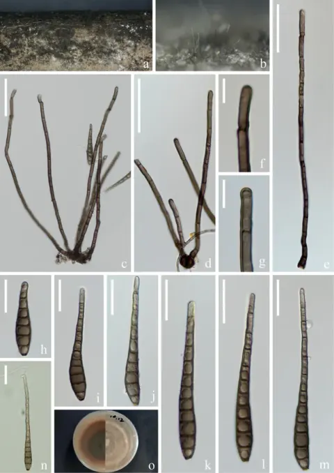
Distoseptispora yunjushanensis (HFJAU 10005, holotype) a, b colonies on bamboo culms c–f conidiophores with conidia g-i young conidia j-l mature conidia m conidium with proliferation n germinating conidium o, p culture on PDA from above and reverse. Scale bars: 100 µm (a, b), 20 µm (c–f, m, n), 5 µm (g–l).
- Jenis
- Pelat ilmiah
- Kredit
- Zhi-Jun Zhai, Jun-Qing Yan, Wei-Wu Li, Yang Gao, Hai-Jing Hu, Jian-Ping Zhou, Hai-Yan Song, Dian-Ming Hu
- Lisensi
- CC BY 4.0
- Sumber media
- Buka entri sumber
Dokumentasi tambahan
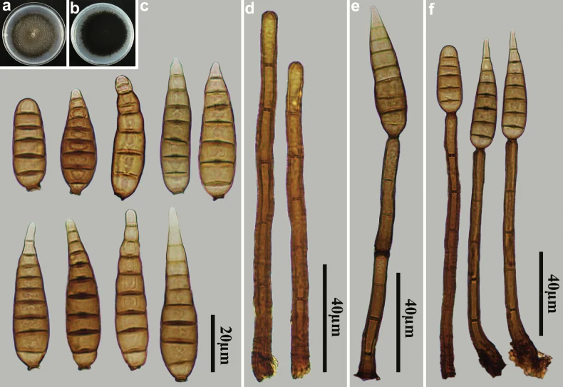
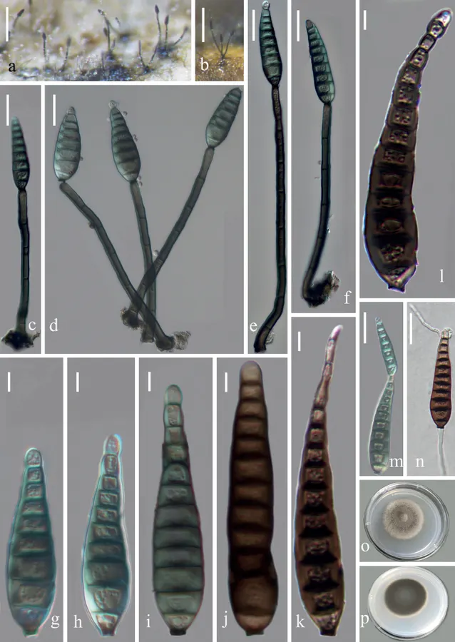
Distoseptispora yunjushanensis (HJAUP M1307) a surface of colony after 4 weeks on PDA b reverse of colony after 4 weeks on PDA c conidia d conidiophores e, f conidiophores, conidiogenous cells and conidia.
Kredit dan penggunaan
Ming-Gen Liao, Xing-Xing Luo, Ya-Fen Hu, Rafael F. Castañeda-Ruíz, Zhao-Huan Xu, Jian Ma
Ukuran dan format gambar disesuaikan tanpa pemotongan, retus, atau penambahan skala. Lisensi sumber dipertahankan.
Sumber profil
- [1] Three novel species of Distoseptispora (Distoseptisporaceae) isolated from bamboo in Jiangxi Province, China — Distoseptispora yunjushanensis
- [2] Catalogue of Life Base2026-09-11 — Distoseptispora yunjushanensis
- [3] Dokumentasi visual — Distoseptispora yunjushanensis
- [4] Dokumentasi visual — Distoseptispora yunjushanensis
Rujukan halaman ini
Fungi. Distoseptispora yunjushanensis. Rilis data fungi-2026-10-12-expansion-2. https://fungi-atlas.mgadib.chatgpt.site/jamur/distoseptispora-yunjushanensis/.
Status: impor sumber; pemeriksaan editorial per profil belum selesai. Untuk klaim biologis, gunakan rujukan pada bagian terkait dan periksa konteks sumber aslinya.