Ciri dan struktur
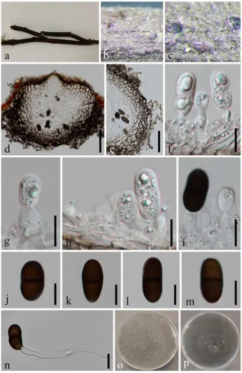
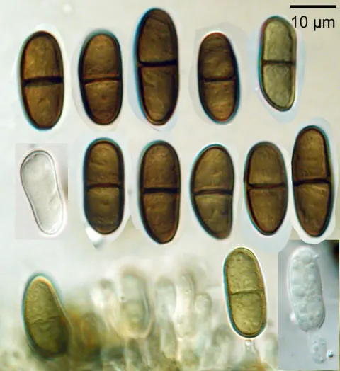
Saprobic on dead twigs of Macadamia sp. Sexual morph: Ascomata 80–120 × 160–220 µm (x̄ = 100 × 193 µm, n = 10), immersed, visible as dark dots on the host surface, under to clypeus, solitary, uni-loculate, ampulliform, papillate, without ostiole. Peridium 50–190 µm wide (x̄ = 95 µm, n = 25), comprising three section layers, the inner section layer composed of hyaline cells of textura angularis, the outer section layer with brown to dark brown cells of textura angularis, and the outermost layer of cells surrounding the ascomata is composed of brown cells of textura prismatica. Hamathecium 4.5–8.5 µm wide (x̄ = 6.5 μm, n = 30), comprising cylindrical, hyaline, septate, cellular pseudoparaphyses. Asci 110–235 × 23–38 µm (x̄ = 173 × 32 µm, n = 30), 6–8-spored, bitunicate, clavate to broadly fusoid-ellipsoid, with furcated pedicel, apically rounded, with a well-developed ocular chamber. Ascospores (27–)30–37(–40) × 14–19 µm (x̄ = 33 × 17 µm, n = 55), overlapping, uniseriate, oval to ellipsoid, hyaline to yellowish brown, aseptate when young, becoming brown to dark brown, 1- or 2- septate at maturity, slightly constricted at the septum, smooth-walled, granular, with mucilaginous polar appendages at one or both ends. Asexual morph: Conidiomata pycnidial produced on PDA within seven weeks, solitary or aggregated, superficial, brown, hairy, globose to subglobose, covered with hyphal hairs, unilocular. Conidiophores reduce to conidiogenous cells. Conidiogenous cells holoblastic, discrete, cylindrical, hyaline, smooth-walled, proliferating percurrently. Conidia 19–26.5 × 10–13.5 µm (x̄ = 22.6 × 11.2 µm, n = 50), hyaline and aseptate when immature, brown to dark brown and one-septate when mature, oblong to ovoid, granular, one end obtuse to slightly rounded ends, one cell slightly wider or same width.
Habitat dan peran ekologis
Saprobic on dead twigs of Macadamia sp. Sexual morph: Ascomata 80–120 × 160–220 µm (x̄ = 100 × 193 µm, n = 10), immersed, visible as dark dots on the host surface, under to clypeus, solitary, uni-loculate, ampulliform, papillate, without ostiole. Peridium 50–190 µm wide (x̄ = 95 µm, n = 25), comprising three section layers, the inner section layer composed of hyaline cells of textura angularis, the outer section layer with brown to dark brown cells of textura angularis, and the outermost layer of cells surrounding the ascomata is composed of brown cells of textura prismatica. Hamathecium 4.5–8.5 µm wide (x̄ = 6.5 μm, n = 30), comprising cylindrical, hyaline, septate, cellular pseudoparaphyses. Asci 110–235 × 23–38 µm (x̄ = 173 × 32 µm, n = 30), 6–8-spored, bitunicate, clavate to broadly fusoid-ellipsoid, with furcated pedicel, apically rounded, with a well-developed ocular chamber. Ascospores (27–)30–37(–40) × 14–19 µm (x̄ = 33 × 17 µm, n = 55), overlapping, uniseriate, oval to ellipsoid, hyaline to yellowish brown, aseptate when young, becoming brown to dark brown, 1- or 2- septate at maturity, slightly constricted at the septum, smooth-walled, granular, with mucilaginous polar appendages at one or both ends.
- Habitat atau konteks temuan
- Informasi belum tersedia pada sumber rilis ini.
- Substrat
- Informasi belum tersedia pada sumber rilis ini.
- Peran atau konteks
- Pengurai
Reproduksi
Saprobic on dead twigs of Macadamia sp. Sexual morph: Ascomata 80–120 × 160–220 µm (x̄ = 100 × 193 µm, n = 10), immersed, visible as dark dots on the host surface, under to clypeus, solitary, uni-loculate, ampulliform, papillate, without ostiole. Peridium 50–190 µm wide (x̄ = 95 µm, n = 25), comprising three section layers, the inner section layer composed of hyaline cells of textura angularis, the outer section layer with brown to dark brown cells of textura angularis, and the outermost layer of cells surrounding the ascomata is composed of brown cells of textura prismatica. Hamathecium 4.5–8.5 µm wide (x̄ = 6.5 μm, n = 30), comprising cylindrical, hyaline, septate, cellular pseudoparaphyses.
Catatan identitas dan cakupan
- Nama diterima mengikuti Catalogue of Life Base2026-09-11; uraian mempertahankan konsep dan nama sumber aslinya.
- Preserve original treatment localities, hosts and substrate scope. Materials examined describe documented specimens, not the full global range.
- Impor dan terjemahan otomatis belum diperiksa editorial per profil. Struktur reproduktif yang disebutkan tidak membuktikan seluruh siklus hidup diketahui.
- Pengukuran pada uraian mengikuti spesimen, kultur, dan metode sumber; angka tidak diperluas ke semua populasi.
Catatan distribusi dalam narasi mengikuti sumbernya. Dokumentasi spesimen atau foto mengikuti catatan sumber. Bukti wilayah Indonesia dan penilaian konservasi tidak disimpulkan dari gambar.
Melihat dengan konteks
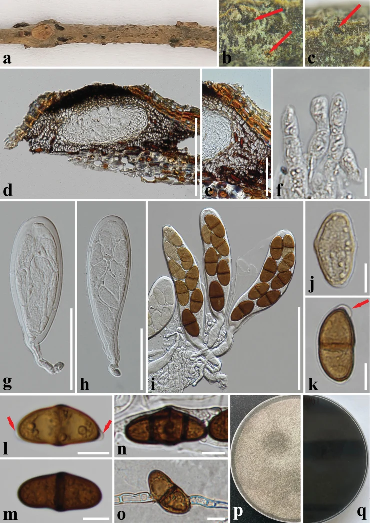
Dothiorella macadamiae (CMUB 40066, holotype) a–c appearance of ascomata on the host surface d vertical section of an ascoma e peridium f hamathecium g–i asci j–n ascospores (arrows indicate mucilaginous polar appendages) o a germinating ascospore p, q colony on PDA (p-front and q-reverse views). Scale bars: 200 µm (d); 100 µm (i); 50 µm (e, g, h); 10 µm (f, j–o).
- Jenis
- Pelat ilmiah
- Kredit
- Xian Zhang, Saowaluck Tibpromma, Samantha C. Karunarathna, Tian-Ye Du, Li-Su Han, Abdallah M. Elgorban, Jaturong Kumla, Chanokned Senwanna, Dong-Qin Dai, Nakarin Suwannarach, Hao-Han Wang
- Lisensi
- CC BY 4.0
- Sumber media
- Buka entri sumber
Dokumentasi tambahan
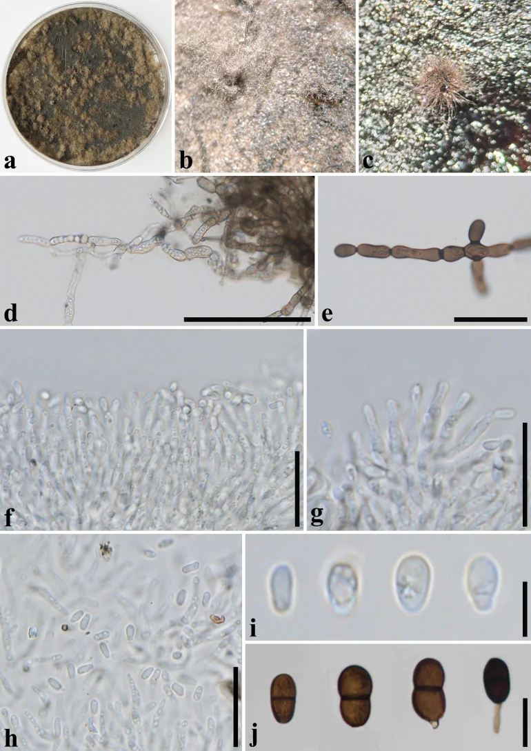
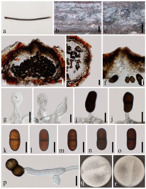
Dothiorella macadamiae (SDBR-CMU512, ex-type) a colony on PDA b, c sporulating colonies on PDA d, e chlamydospores f–h conidiogenous cells with conidia i–j Conidia. Scale bars: 100 µm (d); 50 µm (e); 20 µm (f–h, j); 5 µm (i).
Kredit dan penggunaan
Xian Zhang, Saowaluck Tibpromma, Samantha C. Karunarathna, Tian-Ye Du, Li-Su Han, Abdallah M. Elgorban, Jaturong Kumla, Chanokned Senwanna, Dong-Qin Dai, Nakarin Suwannarach, Hao-Han Wang
Ukuran dan format gambar disesuaikan tanpa pemotongan, retus, atau penambahan skala. Lisensi sumber dipertahankan.
Sumber profil
- [1] Additions to the saprobic fungi (Ascomycota) associated with macadamia trees from the Greater Mekong Subregion — Dothiorella macadamiae
- [2] Catalogue of Life Base2026-09-11 — Dothiorella macadamiae
- [3] Dokumentasi visual — Dothiorella macadamiae
- [4] Dokumentasi visual — Dothiorella macadamiae
Rujukan halaman ini
Fungi. Dothiorella macadamiae. Rilis data fungi-2026-10-12-expansion-2. https://fungi-atlas.mgadib.chatgpt.site/jamur/dothiorella-macadamiae/.
Status: impor sumber; pemeriksaan editorial per profil belum selesai. Untuk klaim biologis, gunakan rujukan pada bagian terkait dan periksa konteks sumber aslinya.