Ciri dan struktur
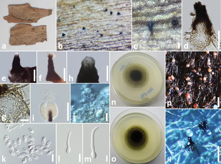
Saprobic on dead leaves of Hedychium coronarium J. Koenig. Sexual morph: Undetermined. Asexual morph: Coelomycetous. Conidiomata 120–140 µm high, 60–70 µm diam., pycnidial, solitary, scattered, superficial, visible as small black spots on host surface, dark brown to black, obpyriform, papillate. Neck up to 80–100 μm long, 20–30 µm diam., elongated, dark brown, usually straight, but sometimes slightly curved. Conidiomatal wall 10–20 µm wide, composed of 4–5 layers of light brown, thick-walled cells, arranged in textura angularis. Conidiophores reduced to conidiogenous cells. Conidiogenous cells 3–4 × 3–3.5 μm (x̄ = 3.6 × 3.2 μm, n = 10), arising from the inner cell wall of the apex, hyaline, aseptate, smooth, ampulliform. Conidia 4–5 × 1.8–2.2 μm (x̄ = 4.6 × 2.1 μm, n = 30), oval to oblong, smooth, thin-walled, hyaline, aseptate, with 1–2-minute guttules. Colonies on PDA reaching 30 mm diameter after 3 weeks at 20–25 °C, colonies medium sparse, circular, raised, surface slightly rough with entire edge, margin entire, colony from above: light brown to grey at the margin, dark brown at middle, dark brown to black at the center; reverse, light brown to yellowish at the margin, brown at middle, dark brown to black at the center; mycelium light brown to grey with tufts; not producing pigments in PDA.
Habitat dan peran ekologis
Saprobic on dead leaves of Hedychium coronarium J. Koenig. Sexual morph: Undetermined. Asexual morph: Coelomycetous. Conidiomata 120–140 µm high, 60–70 µm diam., pycnidial, solitary, scattered, superficial, visible as small black spots on host surface, dark brown to black, obpyriform, papillate. Neck up to 80–100 μm long, 20–30 µm diam., elongated, dark brown, usually straight, but sometimes slightly curved. Conidiomatal wall 10–20 µm wide, composed of 4–5 layers of light brown, thick-walled cells, arranged in textura angularis. Conidiophores reduced to conidiogenous cells. Conidiogenous cells 3–4 × 3–3.5 μm (x̄ = 3.6 × 3.2 μm, n = 10), arising from the inner cell wall of the apex, hyaline, aseptate, smooth, ampulliform. Conidia 4–5 × 1.8–2.2 μm (x̄ = 4.6 × 2.1 μm, n = 30), oval to oblong, smooth, thin-walled, hyaline, aseptate, with 1–2-minute guttules. Colonies on PDA reaching 30 mm diameter after 3 weeks at 20–25 °C, colonies medium sparse, circular, raised, surface slightly rough with entire edge, margin entire, colony from above: light brown to grey at the margin, dark brown at middle, dark brown to black at the center; reverse, light brown to yellowish at the margin, brown at middle, dark brown to black at the center; mycelium light brown to grey with tufts; not producing pigments in PDA.
- Habitat atau konteks temuan
- Informasi belum tersedia pada sumber rilis ini.
- Substrat
- Informasi belum tersedia pada sumber rilis ini.
- Peran atau konteks
- Pengurai
Reproduksi
Saprobic on dead leaves of Hedychium coronarium J. Koenig. Sexual morph: Undetermined. Asexual morph: Coelomycetous. Conidiomata 120–140 µm high, 60–70 µm diam., pycnidial, solitary, scattered, superficial, visible as small black spots on host surface, dark brown to black, obpyriform, papillate. Neck up to 80–100 μm long, 20–30 µm diam., elongated, dark brown, usually straight, but sometimes slightly curved. Conidiomatal wall 10–20 µm wide, composed of 4–5 layers of light brown, thick-walled cells, arranged in textura angularis. Conidiophores reduced to conidiogenous cells. Conidiogenous cells 3–4 × 3–3.5 μm (x̄ = 3.6 × 3.2 μm, n = 10), arising from the inner cell wall of the apex, hyaline, aseptate, smooth, ampulliform.
Catatan identitas dan cakupan
- Nama diterima mengikuti Catalogue of Life Base2026-09-11; uraian mempertahankan konsep dan nama sumber aslinya.
- Preserve original treatment localities, hosts and substrate scope. Materials examined describe documented specimens, not the full global range.
- Impor dan terjemahan otomatis belum diperiksa editorial per profil. Struktur reproduktif yang disebutkan tidak membuktikan seluruh siklus hidup diketahui.
- Pengukuran pada uraian mengikuti spesimen, kultur, dan metode sumber; angka tidak diperluas ke semua populasi.
Catatan distribusi dalam narasi mengikuti sumbernya. Dokumentasi spesimen atau foto mengikuti catatan sumber. Bukti wilayah Indonesia dan penilaian konservasi tidak disimpulkan dari gambar.
Melihat dengan konteks
Elongaticollum hedychii (MFLU 18-2542, holotype) a specimen b appearance of conidiomata on host c close up of conidiomata on host d vertical section through conidioma e, f squash mount of conidioma g conidioma wall h, i elongated conidiomatal necks j conidiogenous cells k conidia l, m germinated conidia n colony from below o colony from above p, q pycnidia formed on PDA. Scale bars: 100 µm (c), 50 µm (d–h), 10 µm (g), 30 µm (i), 3 µm (j–m), 100 µm (p, q).
- Jenis
- Pelat ilmiah
- Kredit
- Danushka S. Tennakoon, Kasun M. Thambugala, Dhanushka N. Wanasinghe, Eleni Gentekaki, Itthayakorn Promputtha, Chang- Hsin Kuo, Kevin D. Hyde
- Lisensi
- CC BY 4.0
- Sumber media
- Buka entri sumber
Sumber profil
- [1] Additions to Phaeosphaeriaceae (Pleosporales): Elongaticollum gen. nov., Ophiosphaerella taiwanensis sp. nov., Phaeosphaeriopsis beaucarneae sp. nov. and a new host record of Neosetophoma poaceicola from Musaceae — Elongaticollum hedychii
- [2] Catalogue of Life Base2026-09-11 — Elongaticollum hedychii
- [3] Dokumentasi visual — Elongaticollum hedychii
Rujukan halaman ini
Fungi. Elongaticollum hedychii. Rilis data fungi-2026-10-12-expansion-2. https://fungi-atlas.mgadib.chatgpt.site/jamur/elongaticollum-hedychii/.
Status: impor sumber; pemeriksaan editorial per profil belum selesai. Untuk klaim biologis, gunakan rujukan pada bagian terkait dan periksa konteks sumber aslinya.