Ciri dan struktur
Sexual form: unknown. Asexual form: Hyalorhinocladiella -like. Conidiophores mononematous, micronematous; conidiophorous cells solitary, integrated, flask-shaped, with an inflated base (3.6–) 4.6–6.1 (–7.1) μm in diam., the fertile hyphoid part (9.1–) 12.2–19.0 (–22.5) × (1.4–) 1.9–3.1 (–4.7) μm, often crooked due to successive conidial development; conidia 1-celled, asymmetrically ellipsoidal in face view, concave, lunate in side view, with a layer of adhesive mucus on the concave surface, ending slightly apiculate, hyaline, smooth, (8.0–) 10–12 (–13.1) × (3.3–) 3.4–4.5 (–5.1) μm, containing an ovoid endospore-like structure. Colonies on 2% MEA in the dark reaching 31 mm in diam. in 8 days at 25 °C, growth rate up to 5 mm/day at the fastest; colony margin smooth. Mycelium compact, somewhat floccose in the margin, white at first, gradually discolouring to greyish-green, eventually dark green. Optimal growth temperature 25 °C, growth at 5 °C and 35 °C.
Habitat dan peran ekologis
Esteya vermicola is known only from an asexual, Hyalorhinocladiella -like state producing lunate and bacilliform conidia (Liou et al. 1999, Kubátová et al. 2000, Wang et al. 2009, 2014) that we also observed in various strains of E. vermicola with a different origin (Taiwan, Korea, Czech Republic). Our strain was identified as E. vermicola based on phylogenetic inferences and morphological characters. However, our strain differed from previous descriptions (Liou et al. 1999) in having only lunate conidia in vitro. The size of the lunate conidia of our strains (mostly 10 - 12 × 3.4 - 4.5 μm) was similar to that reported for E. vermicola, viz. 9.9–11.9 × 3.4–4.5 μm vs 8.2-11.1 × 3.5–3.7 μm (Taiwan, Liou et al. 1999), 9.3–12.4 × 3.0–3.2 μm (Czech Republic, Kubátová et al. 2000), 7.7–12.1 × 3.0–3.8 μm (Korea, Wang et al. 2009) or 8.7–11.9 × 3.0–3.6 μm (Brazil, Wang et al. 2014). This is the first report of E. vermicola from continental China. The species was originally isolated from Japanese black pine infected by the pinewood nematode Bursaphelenchus xylophilus, in Taiwan (Liou et al. 1999). Since then, its distribution range has been extended to Japan and Korea, Europe (Czech Republic, Italy) and both North (USA) and South America (Brazil) (Liou et al. 1999, Kubátová et al. 2000, Wang et al. 2009, 2014, Li et al. 2018).
- Habitat atau konteks temuan
- Informasi belum tersedia pada sumber rilis ini.
- Substrat
- Informasi belum tersedia pada sumber rilis ini.
- Peran atau konteks
- Belum dikategorikan pada rilis ini.
Reproduksi
Sexual form: unknown. Asexual form: Hyalorhinocladiella -like. Conidiophores mononematous, micronematous; conidiophorous cells solitary, integrated, flask-shaped, with an inflated base (3.6–) 4.6–6.1 (–7.1) μm in diam., the fertile hyphoid part (9.1–) 12.2–19.0 (–22.5) × (1.4–) 1.9–3.1 (–4.7) μm, often crooked due to successive conidial development; conidia 1-celled, asymmetrically ellipsoidal in face view, concave, lunate in side view, with a layer of adhesive mucus on the concave surface, ending slightly apiculate, hyaline, smooth, (8.0–) 10–12 (–13.1) × (3.3–) 3.4–4.5 (–5.1) μm, containing an ovoid endospore-like structure.
Catatan identitas dan cakupan
- Nama diterima mengikuti Catalogue of Life Base2026-09-11; uraian mempertahankan konsep dan nama sumber aslinya.
- Preserve original treatment localities, hosts and substrate scope. Materials examined describe documented specimens, not the full global range.
- Impor dan terjemahan otomatis belum diperiksa editorial per profil. Struktur reproduktif yang disebutkan tidak membuktikan seluruh siklus hidup diketahui.
- Pengukuran pada uraian mengikuti spesimen, kultur, dan metode sumber; angka tidak diperluas ke semua populasi.
Catatan distribusi dalam narasi mengikuti sumbernya. Dokumentasi spesimen atau foto mengikuti catatan sumber. Bukti wilayah Indonesia dan penilaian konservasi tidak disimpulkan dari gambar.
Melihat dengan konteks
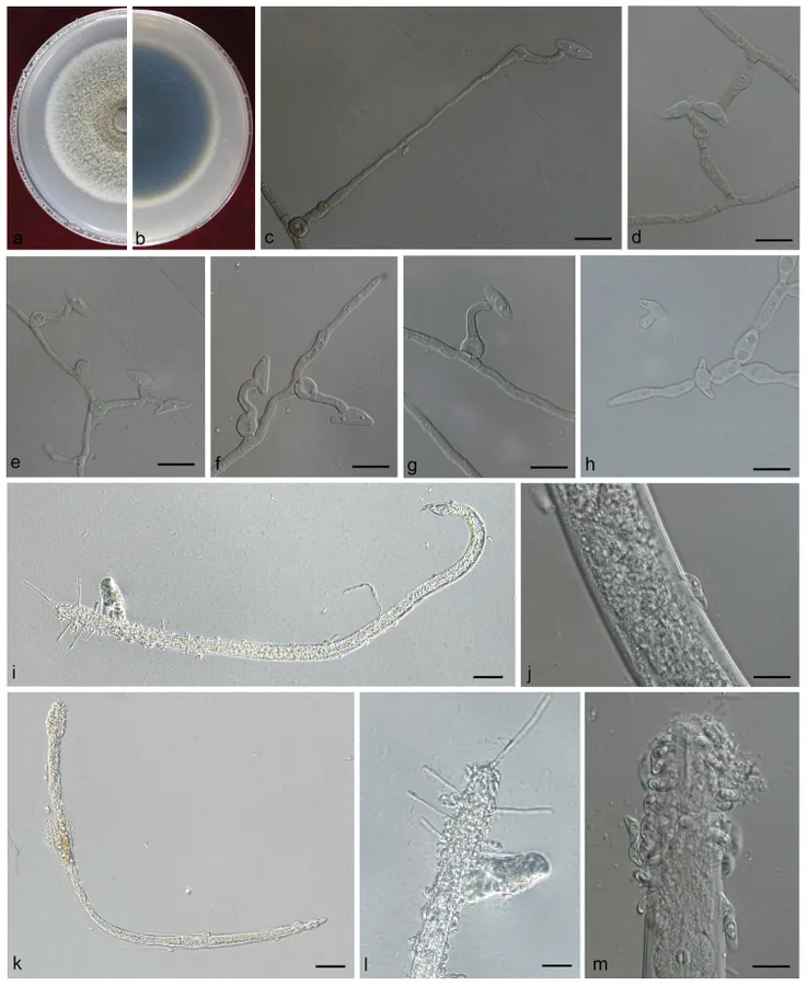
A–H Morphological characters of Esteya vermicola A, B upper and reverse of cultures on 2% MEA 20 d after inoculation C–H conidiogenous cells with lunate conidia I–M the cuticle of a nematode attached by many lunate conidia. Scale bars: 20 μm (I, K, L); 10 μm (C–H, J, M).
- Jenis
- Pelat ilmiah
- Kredit
- Wang HuiMin, Zheng Wang, Fu Liu, Cheng Xu Wu, Su Fang Zhang, Xiang Bo Kong, Cony Decock, Quan Lu, Zhen Zhang
- Lisensi
- CC BY 4.0
- Sumber media
- Buka entri sumber
Sumber profil
- [1] Differential patterns of ophiostomatoid fungal communities associated with three sympatric Tomicus species infesting pines in south-western China, with a description of four new species — Esteya vermicola
- [2] Catalogue of Life Base2026-09-11 — Esteya vermicola
- [3] Dokumentasi visual — Esteya vermicola
Rujukan halaman ini
Fungi. Esteya vermicola. Rilis data fungi-2026-10-12-expansion-2. https://fungi-atlas.mgadib.chatgpt.site/jamur/esteya-vermicola/.
Status: impor sumber; pemeriksaan editorial per profil belum selesai. Untuk klaim biologis, gunakan rujukan pada bagian terkait dan periksa konteks sumber aslinya.