Ciri dan struktur
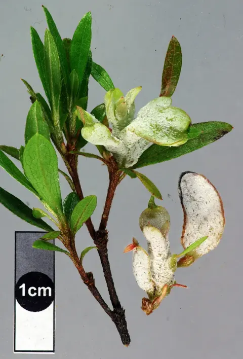
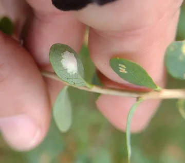
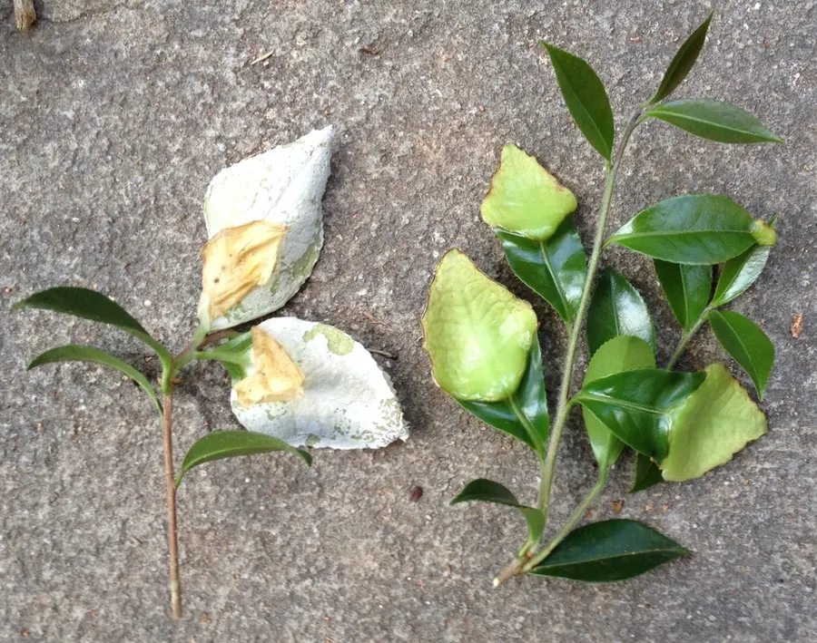
Exobasidium camelliae is a phytopathogenic fungus that infects ornamental shrubs of the Camellia genus. It absorbs nutrients from the host through its haustoria and causes the leaves of the host plant to be thicker and lighter green than usual. It forms a hymenium between cells four to six layers above the lower epidermis which is subsequently sloughed off to reveal its basidia. The hyphae of E. camelliae grow in the intercellular spaces of the host plant’s cells. The hyphae are thick walled and septate. The septa can be either true septa or pseudosepta. True septa are single small pores between cells which have pore plugs that control the flow of material through the fungus. Pseuduosepta are single large pores that allow the movement of larger materials and organelles through the fungus. Pseudosepta do not have pore plugs and form through the invagination of entire lateral walls. In contrast to other basidiomycetes, E. Camelliae do not form clamp connections. The haustoria of E. camelliae are projections into host cells walls with slender, branched, fingerlike lobes. A collar of host cell wall material remains at each lobe’s entry point into the cell, and host cells can synthesize cell wall material to surround the lobes as a form of protection. The haustoria contain inclusion bodies which appear in the extracellular space between the lobe’s plasma membrane and cell wall. These inclusion bodies give rise to the haustorial cap which acts as a barrier between the lobes and the hyphae. While the lobes contain similar cytoplasmic material to the hyphae, they do not contain nuclei.
Habitat dan peran ekologis
The hyphae of E. camelliae grow in the intercellular spaces of the host plant’s cells. The hyphae are thick walled and septate. The septa can be either true septa or pseudosepta. True septa are single small pores between cells which have pore plugs that control the flow of material through the fungus. Pseuduosepta are single large pores that allow the movement of larger materials and organelles through the fungus. Pseudosepta do not have pore plugs and form through the invagination of entire lateral walls. In contrast to other basidiomycetes, E. Camelliae do not form clamp connections. Species of the Cladosporium genus have been observed to grow on the hymenium of E. camelliae. The Cladiosporium grow on top of the exposed basidia, lysing the cells and inducing the release of their contents. This is in contrast to other Cladiosporium relationships where the parasite grows inside the cells of their host. It is still unclear whether this is a parasitic, commensal or mutualistic relationship.
- Habitat atau konteks temuan
- Informasi belum tersedia pada sumber rilis ini.
- Substrat
- Informasi belum tersedia pada sumber rilis ini.
- Peran atau konteks
- Belum dikategorikan pada rilis ini.
Reproduksi
Exobasidium camelliae is a phytopathogenic fungus that infects ornamental shrubs of the Camellia genus. It absorbs nutrients from the host through its haustoria and causes the leaves of the host plant to be thicker and lighter green than usual. It forms a hymenium between cells four to six layers above the lower epidermis which is subsequently sloughed off to reveal its basidia. The hyphae of E. camelliae grow in the intercellular spaces of the host plant’s cells. The hyphae are thick walled and septate. The septa can be either true septa or pseudosepta. True septa are single small pores between cells which have pore plugs that control the flow of material through the fungus. Pseuduosepta are single large pores that allow the movement of larger materials and organelles through the fungus.
Catatan identitas dan cakupan
- Nama diterima mengikuti Catalogue of Life Base2026-09-11; uraian mempertahankan konsep dan nama sumber aslinya.
- Original geographic, specimen and life-cycle scope retained; source import awaits individual editorial review.
- Impor dan terjemahan otomatis belum diperiksa editorial per profil. Struktur reproduktif yang disebutkan tidak membuktikan seluruh siklus hidup diketahui.
- Pengukuran pada uraian mengikuti spesimen, kultur, dan metode sumber; angka tidak diperluas ke semua populasi.
Catatan distribusi dalam narasi mengikuti sumbernya. Dokumentasi spesimen atau foto mengikuti catatan sumber. Bukti wilayah Indonesia dan penilaian konservasi tidak disimpulkan dari gambar.
Melihat dengan konteks
Dokumentasi Exobasidium camelliae menurut asosiasi foto pada record takson iNaturalist. Kredit: Henry Van T. Cotter. Identitas mengikuti sumber; bukan penentuan spesimen independen.
- Jenis
- Fotografi
- Kredit
- (c) Henry Van T. Cotter, some rights reserved (CC BY), uploaded by Henry Van T. Cotter
- Lisensi
- CC BY 4.0
- Sumber media
- Buka entri sumber
Sumber profil
- [1] Exobasidium camelliae — Wikipedia (en)
- [2] Catalogue of Life Base2026-09-11 — Exobasidium camelliae
- [3] Dokumentasi visual — Exobasidium camelliae
Rujukan halaman ini
Fungi. Exobasidium camelliae. Rilis data fungi-2026-10-12-expansion-2. https://fungi-atlas.mgadib.chatgpt.site/jamur/exobasidium-camelliae/.
Status: impor sumber; pemeriksaan editorial per profil belum selesai. Untuk klaim biologis, gunakan rujukan pada bagian terkait dan periksa konteks sumber aslinya.