Ciri dan struktur
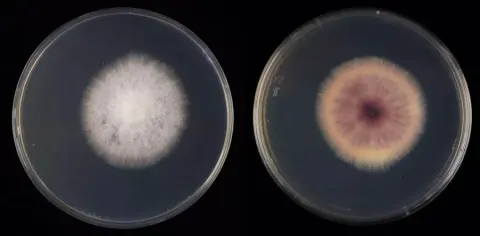
Different from F. thapsinum by the absence of napiform microconidia. Different from F. andiyazi, F. thapsinum and F. verticillioides by its lighter colony pigmentation, growth rates, microconidial septation, presence of true chlamydospores and secondary metabolite patterns. Colonies grown in the dark at 24°C. On MEA and PDA with an average radial growth rate of 5.9–6.5 mm/d and filling an entire 90 mm Petri dish in 7 d. Surface white to pale rosy buff, flat, velvety to felty with abundant patches of white aerial mycelium; margin regular, filiform. Reverse pale saffron to peach, a pale bay diffusible pigment can be scarcely produced. On OA, occupying an entire 90 mm Petri dish in 7 d. Surface white to pale rosy buff, flat, velvety to felty with abundant patches of white aerial mycelium; margin regular. Reverse pale luteous to saffron. On SNA, reaching 24–25 mm diam. in 7 d. Surface white, velvety, with scarce aerial mycelium, margins filiform. Reverse white. Conidiophores on aerial mycelium straight, erect, septate, smooth- and thin-walled, commonly simple or reduced to conidiogenous cells, borne laterally on hyphae or laterally branched at various levels, bearing terminal single monophialides; phialides subulate to subcylindrical, smooth- and thin-walled, (17–)25.5–39.5 μm long, (2–)2.5–3.5 μm at widest point, periclinal thickening and collarettes inconspicuous or absent; microconidia hyaline, clavate, smooth- and thin-walled, 0–3-septate, (7–)9–15(–21) × (2–)2.5–4(–5) μm, arranged in long chains at the tip of monophialides. Sporodochia pale to bright orange, formed abundantly on the surface of carnation leaves and on agar surface.
Habitat dan peran ekologis
Although clearly recognisable based on genetic markers, Fusarium madaense is hardly distinguishable from its closer relatives, based on morphological features only. The novel species is characterised by abundant long, slender and slightly curved macroconidia, a morphology typical of the FFSC. The overall morphology of F. madaense is similar to that of F. andiyazi, F. thapsinum and F. verticillioides; all species are characterised by clavate microconidia formed in long chains from relatively long monophialides. Moreover, the four mentioned species are known to be pathogenic on sorghum (Leslie and Summerell 2006) and have been isolated here from the same geographical regions. Nevertheless, some morphological features of F. madaense can provide an indication of its identity. These include a pale saffron colony pigmentation on OA and PDA, not developing the intense purple colour typical of F. andiyazi and F. verticillioides, nor the yellow pigmentation of F. thapsinum; the presence of up to 3-septate clavate microconidia vs. the up to 2-septate and aseptate microconidia of F. andiyazi and F. verticillioides, respectively; and the aseptate, but also rarely napiform microconidia of F. thapsinum (Nirenberg 1976, Klittich et al. 1997, Marasas et al. 2001).
- Habitat atau konteks temuan
- Informasi belum tersedia pada sumber rilis ini.
- Substrat
- Informasi belum tersedia pada sumber rilis ini.
- Peran atau konteks
- Belum dikategorikan pada rilis ini.
Reproduksi
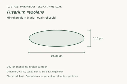
Colonies grown in the dark at 24°C. On MEA and PDA with an average radial growth rate of 5.9–6.5 mm/d and filling an entire 90 mm Petri dish in 7 d. Surface white to pale rosy buff, flat, velvety to felty with abundant patches of white aerial mycelium; margin regular, filiform. Reverse pale saffron to peach, a pale bay diffusible pigment can be scarcely produced. On OA, occupying an entire 90 mm Petri dish in 7 d. Surface white to pale rosy buff, flat, velvety to felty with abundant patches of white aerial mycelium; margin regular. Reverse pale luteous to saffron. On SNA, reaching 24–25 mm diam. in 7 d. Surface white, velvety, with scarce aerial mycelium, margins filiform. Reverse white. Conidiophores on aerial mycelium straight, erect, septate, smooth- and thin-walled, commonly simple or reduced to conidiogenous cells, borne laterally on hyphae or laterally branched at various levels, bearing terminal single monophialides; phialides subulate to subcylindrical, smooth- and thin-walled, (17–)25.5–39.5 μm long, (2–)2.5–3.5 μm at widest point, periclinal thickening and collarettes inconspicuous or absent; microconidia hyaline, clavate, smooth- and thin-walled, 0–3-septate, (7–)9–15(–21) × (2–)2.5–4(–5) μm, arranged in long chains at the tip of monophialides.
Catatan identitas dan cakupan
- Nama diterima mengikuti Catalogue of Life Base2026-09-11; uraian mempertahankan konsep dan nama sumber aslinya.
- Preserve original treatment localities, hosts and substrate scope. Materials examined describe documented specimens, not the full global range.
- Impor dan terjemahan otomatis belum diperiksa editorial per profil. Struktur reproduktif yang disebutkan tidak membuktikan seluruh siklus hidup diketahui.
- Pengukuran pada uraian mengikuti spesimen, kultur, dan metode sumber; angka tidak diperluas ke semua populasi.
Catatan distribusi dalam narasi mengikuti sumbernya. Dokumentasi spesimen atau foto mengikuti catatan sumber. Bukti wilayah Indonesia dan penilaian konservasi tidak disimpulkan dari gambar.
Melihat dengan konteks
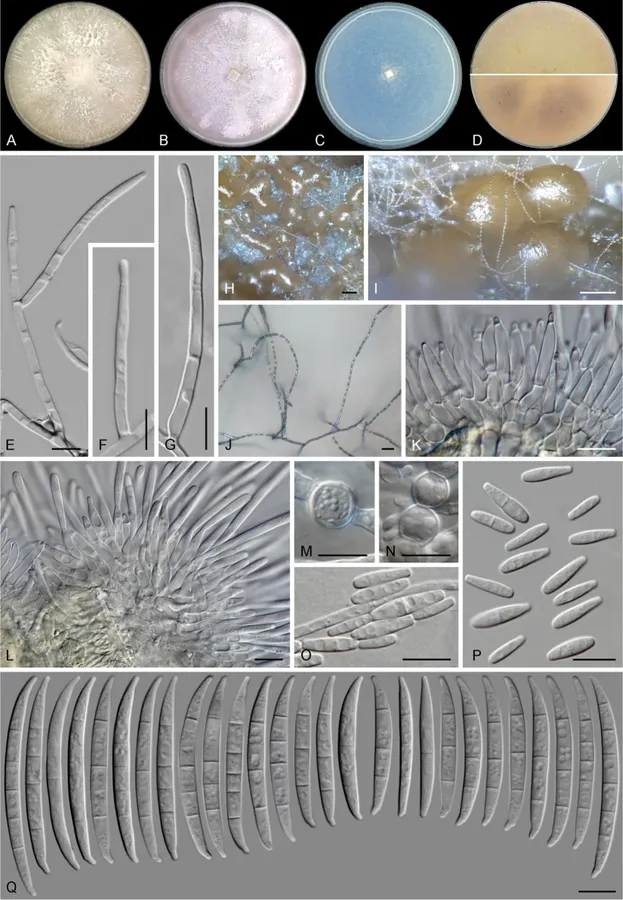
Fusarium madaense sp. nov. (ex-type culture CBS 146669). A–C aspect of colonies on PDA, OA and SNA, respectively, after 14 d at 24°C in the dark D colony reverse on OA (up) and PDA (down) after 14 d at 24 °C in the dark E–G, J aerial conidiophores and phialides H, I sporodochia formed on the surface of carnation leaves K, L sporodochial conidiophores M, N chlamydospores O, P microconidia Q sporodochial conidia. Scale bars: 100 μm (H, I); 20 μm (J); 10 μm (all others).
- Jenis
- Pelat ilmiah
- Kredit
- Chibundu N. Ezekiel, Bart Kraak, Marcelo Sandoval-Denis, Michael Sulyok, Oluwawapelumi A. Oyedele, Kolawole I. Ayeni, Oluwadamilola M. Makinde, Oluwatosin M. Akinyemi, Rudolf Krska, Pedro W. Crous, Jos Houbraken
- Lisensi
- CC BY 4.0
- Sumber media
- Buka entri sumber
Sumber profil
- [1] Diversity and toxigenicity of fungi and description of Fusarium madaense sp. nov. from cereals, legumes and soils in north-central Nigeria — Fusarium madaense
- [2] Catalogue of Life Base2026-09-11 — Fusarium madaense
- [3] Dokumentasi visual — Fusarium madaense
Rujukan halaman ini
Fungi. Fusarium madaense. Rilis data fungi-2026-10-12-expansion-2. https://fungi-atlas.mgadib.chatgpt.site/jamur/fusarium-madaense/.
Status: impor sumber; pemeriksaan editorial per profil belum selesai. Untuk klaim biologis, gunakan rujukan pada bagian terkait dan periksa konteks sumber aslinya.