Ciri dan struktur
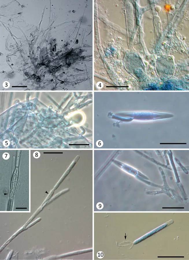
Thalli measuring up to 600 μm long. Basal cell broadly inflated (18–30 μm diam.) and often branched (Fig. 3), bearing a small discoid secreted holdfast at the base or laterally to the basal cell axis (Fig. 4). Dichotomous branching above the basal cell; distal branches bearing spores (Figs 2, 8). Trichospores cylindrical, with a terminal refractive cap (not always visible), measuring 44–56 × 4.5–5.5 μm, with 3 appendages, one central long filiform appendage, coiled around two shorter (about 15–20 μm) and broader lateral appendages (Fig. 10). These appendages can be seen within the generative cell while still attached (Figs 7, 8 arrowhead). Fertile branches bearing 3–4 generative cells, measuring 20–35 × 4–6 μm. Zygospores biconical, 48–60 × 7.5–9.5 μm, with a collar 5–10 (–16) × 4 μm, attached eccentrically and laterally (Type II) to a zygosporophore 20–30 μm long, arising from the conjugation tube in series of scalariform conjugations (Fig. 9). In the hindgut of Baetidae (Ephemeroptera) nymphs.
Habitat dan peran ekologis
The genus Glotzia has nine species (including that described here), all of them sharing the characteristic cylindrical trichospores with a slightly globose cap and the peculiar 3-appendage arrangement observed also in G. balkanensis. This new species mostly resembles the type species G. centroptili described by Gauthier (1936) in French pools and streams of Dauphiné province (south-eastern France) from Centroptilum luteolum nymphs (Baetidae). This French species was recently rediscovered in Catalonia (Spain) also within Centroptilum sp. nymphs (Busquets et al. 2018). This second observation in Spain was important for providing new material to complete the original description, which was scant and had no photographs, only a drawing of a single specimen (Gauthier 1936). The specimens from Bulgaria can be differentiated from G. centroptili by spore characteristics. Trichospores of the Bulgarian species are longer than those observed in France or Spain (40 × 4 μm according to Gauthier (1936); 35–43 × 4–6 μm according to Busquets et al. (2018), up to 56 μm in the specimens reported here). All the fertile branches observed had a maximum of four generative cells in G. balkanensis, while up to seven have been reported in G. centroptili.
- Habitat atau konteks temuan
- Informasi belum tersedia pada sumber rilis ini.
- Substrat
- Informasi belum tersedia pada sumber rilis ini.
- Peran atau konteks
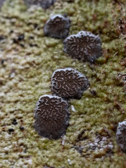
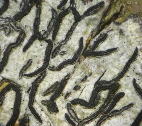
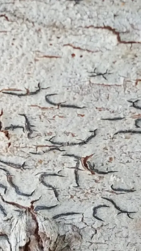
- Pembentuk liken
Reproduksi
Thalli measuring up to 600 μm long. Basal cell broadly inflated (18–30 μm diam.) and often branched (Fig. 3), bearing a small discoid secreted holdfast at the base or laterally to the basal cell axis (Fig. 4). Dichotomous branching above the basal cell; distal branches bearing spores (Figs 2, 8). Trichospores cylindrical, with a terminal refractive cap (not always visible), measuring 44–56 × 4.5–5.5 μm, with 3 appendages, one central long filiform appendage, coiled around two shorter (about 15–20 μm) and broader lateral appendages (Fig. 10). These appendages can be seen within the generative cell while still attached (Figs 7, 8 arrowhead). Fertile branches bearing 3–4 generative cells, measuring 20–35 × 4–6 μm.
Catatan identitas dan cakupan
- Nama diterima mengikuti Catalogue of Life Base2026-09-11; uraian mempertahankan konsep dan nama sumber aslinya.
- Preserve original treatment localities, hosts and substrate scope. Materials examined describe documented specimens, not the full global range.
- Impor dan terjemahan otomatis belum diperiksa editorial per profil. Struktur reproduktif yang disebutkan tidak membuktikan seluruh siklus hidup diketahui.
- Pengukuran pada uraian mengikuti spesimen, kultur, dan metode sumber; angka tidak diperluas ke semua populasi.
Catatan distribusi dalam narasi mengikuti sumbernya. Dokumentasi spesimen atau foto mengikuti catatan sumber. Bukti wilayah Indonesia dan penilaian konservasi tidak disimpulkan dari gambar.
Melihat dengan konteks
Glotzia balkanensis sp. nov. from Baetidae nymphs. 3 thallus overview, note the inflated and branched basal cell structures 4, 5 detail of various swollen basal cells 6 loose zygospore with a collar 7, 8 trichospores on fertile branches, see the appendages inside the generative cell (arrowheads) 9 Zygospore arising from a conjugation tube 10 loose trichospore with a central long filiform appendage and two smaller lateral appendages. Scale bars: 50 µm (3), 25 µm (4–10).
- Jenis
- Pelat ilmiah
- Kredit
- Laia Guàrdia Valle, Desislava Stoianova
- Lisensi
- CC BY 4.0
- Sumber media
- Buka entri sumber
Sumber profil
- [1] First record of Harpellales, Orphellales (Kickxellomycotina) and Amoebidiales (Mesomycetozoea) from Bulgaria, including a new species of Glotzia — Glotzia balkanensis
- [2] Catalogue of Life Base2026-09-11 — Glotzia balkanensis
- [3] Dokumentasi visual — Glotzia balkanensis
Rujukan halaman ini
Fungi. Glotzia balkanensis. Rilis data fungi-2026-10-12-expansion-2. https://fungi-atlas.mgadib.chatgpt.site/jamur/glotzia-balkanensis/.
Status: impor sumber; pemeriksaan editorial per profil belum selesai. Untuk klaim biologis, gunakan rujukan pada bagian terkait dan periksa konteks sumber aslinya.