01 / Morfologi

Ciri dan struktur

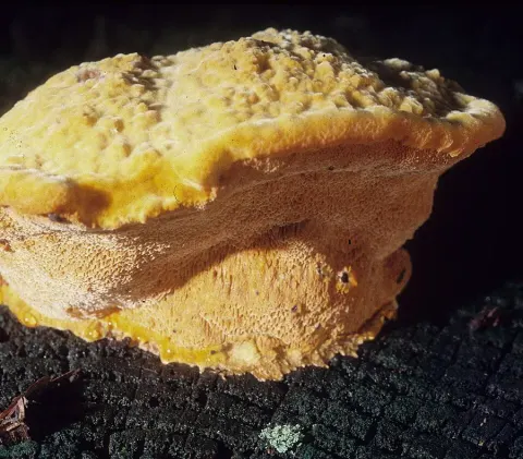
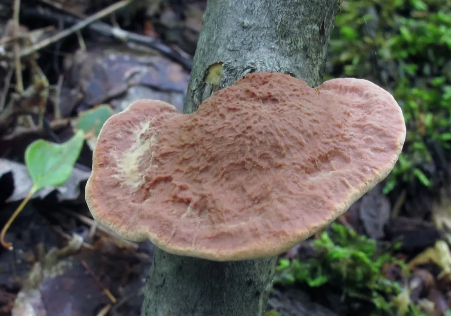
The cap is fan-shaped to semicircular and convex, reaching a diameter of 2.5 – 12 cm. It lacks a stalk, and is instead attached broadly to the substrate. The fruit bodies are initially soft and spongy, but become hard and brittle once they have lost their moisture. The cap surface is covered with matted hairs, has shallow concentric furrows, and a dull brownish-orange color. The flesh is up to 3 cm thick at the thickest part, and is a pale cinnamon color. The pore surface is yellowish to brownish, and the pores are angular, numbering about 2–4 per millimeter. In young fruit bodies, the pore surface bruises reddish brown. The mushroom's odor and taste ranges from sweetish to indistinct. Fruit bodies produce a white spore print. Spores are elliptical to cylindrical, smooth, hyaline (translucent), and measure 3.5–5 by 2–3 μm. The basidia (spore-bearing cells) are club shaped and four-spored, with dimensions of 18–22 by 5–5.6 μm. H. rutilans has a monomitic hyphal system, containing only generative hyphae with clamp connections. In the flesh, these hyphae are thick-walled and highly branched, measuring up to 10 μm in diameter; hyphae comprising the pores and the subhymenium are thinner (up to 6 μm wide) and less branched. A chemical test can be used to help identify H. rutilans: all parts of the fruit body will instantly stain bright violet if a drop of an alkaline solution is applied. Dilute (3–10%) potassium hydroxide (KOH), is often used for this purpose.

Sebagian uraian ditampilkan dalam bahasa sumber; terjemahannya belum tersedia atau belum lolos pemeriksaan.

Rujukan: [1]

Pengukuran mengikuti objek, spesimen atau kultur, dan metode yang disebutkan sumber. Baca uraian asli untuk konteks lengkap.

02 / Kehidupan

Habitat dan peran ekologis

A saprobic species, Hapalopilus rutilans causes a white rot in its host. Fruit bodies of the fungus grow singly, in groups, or in fused and overlapping clusters on the wood of dead and decaying deciduous trees. Preferred hosts include Quercus (oak), Fagus (beech), and Betula (birch), although on rare occasions it has been recorded on conifer wood as well. In central Europe, its preferred host is oak, while in northern Europe it is found most commonly on Corylus and Sorbus. Fruiting typically occurs from early summer to autumn, but the tough fruit bodies are persistent and may be encountered out of the usual growing season. Hapalopilus rutilans has a mostly circumboreal distribution in the north temperate zone, and has been found in North Africa, Asia, Europe, and North America. Outside of this region, it has been recorded from Australia, and Oceania. In North America, where it can be found as far north as the Northwest Territories in Canada, it is more common in the eastern and southwestern part of the continent. In Europe, its northern distribution extends to Porsanger in Norway. Reported for the first time from India in 2011, it was found in forests depots of Chhattisgarh, growing on the stored logs of several native trees: Anogeissus latifolia, Chloroxylon swietenia, Desmodium oojeinense, Shorea robusta, and Terminalia elliptica.

Sebagian uraian ditampilkan dalam bahasa sumber; terjemahannya belum tersedia atau belum lolos pemeriksaan.

Rujukan: [1]

Habitat atau konteks temuan
hutan
Substrat
Informasi belum tersedia pada sumber rilis ini.
Peran atau konteks
Pengurai
Temukan hubungan serupa
03 / Siklus hidup

Reproduksi

Fruit bodies produce a white spore print. Spores are elliptical to cylindrical, smooth, hyaline (translucent), and measure 3.5–5 by 2–3 μm. The basidia (spore-bearing cells) are club shaped and four-spored, with dimensions of 18–22 by 5–5.6 μm. H. rutilans has a monomitic hyphal system, containing only generative hyphae with clamp connections. In the flesh, these hyphae are thick-walled and highly branched, measuring up to 10 μm in diameter; hyphae comprising the pores and the subhymenium are thinner (up to 6 μm wide) and less branched. A chemical test can be used to help identify H. rutilans: all parts of the fruit body will instantly stain bright violet if a drop of an alkaline solution is applied.

Sebagian uraian ditampilkan dalam bahasa sumber; terjemahannya belum tersedia atau belum lolos pemeriksaan.

Rujukan: [1]

Sinonim menurut acuan taksonomi rilis: Agaricus nidulans; Boletus nidulans; Boletus resupinatus; Boletus rutilans; Cyphella rutilans; Fomes resupinatus; Fomes spongiosus; Hapalopilus nidulans; Hemidiscia rutilans; Inodermus rutilans; Inonotus nidulans; Inonotus rutilans; Leptoporus rutilans; Phaeolus nidulans; Phaeolus rutilans; Phaeolus rutilans f. abietis-sibiricae; Phaeolus rutilans f. porioides; Phaeolus rutilans f. resupinatus; Polyporus nidulans; Polyporus nidulans subsp. spongiosus; Polyporus nidulans var. spongiosus; Polyporus rutilans; Polyporus rutilans var. nidulans; Polyporus spongiosus; Polystictus nidulans; Polystictus rutilans; Poria resupinata; Poria spongiosa; Scindalma spongiosum; Trametes lignicola var. populina
04 / Batas informasi

Catatan identitas dan cakupan

  • Nama diterima mengikuti Catalogue of Life Base2026-09-11; uraian mempertahankan konsep dan nama sumber aslinya.
  • Original geographic, specimen and life-cycle scope retained; source import awaits individual editorial review.
  • Impor dan terjemahan otomatis belum diperiksa editorial per profil. Struktur reproduktif yang disebutkan tidak membuktikan seluruh siklus hidup diketahui.
  • Pengukuran pada uraian mengikuti spesimen, kultur, dan metode sumber; angka tidak diperluas ke semua populasi.

Catatan distribusi dalam narasi mengikuti sumbernya. Dokumentasi spesimen atau foto mengikuti catatan sumber. Bukti wilayah Indonesia dan penilaian konservasi tidak disimpulkan dari gambar.

05 / Dokumentasi

Melihat dengan konteks

Dokumentasi Hapalopilus rutilans menurut asosiasi foto pada record takson iNaturalist. Kredit: mycowalt. Identitas mengikuti sumber; bukan penentuan spesimen independen.

Jenis
Fotografi
Kredit
(c) mycowalt, some rights reserved (CC BY-SA), uploaded by mycowalt
Sumber media
Buka entri sumber

Gambar menunjukkan objek menurut identifikasi sumber, bukan verifikasi independen spesimen oleh Fungi. Tidak ada skala baru yang ditambahkan.

06 / Rujukan

Sumber profil

  1. [1] Hapalopilus rutilans — Wikipedia (en)Bagian sumber: Frozen revision 1315525682
    Tanggal penghimpunan sumber: 2026-10-12
    Teks sumber: CC BY-SA4.0 · Wikipedia contributors
  2. [2] Catalogue of Life Base2026-09-11 — Hapalopilus rutilansBagian sumber: ColDP NameUsage.tsv ID 3JKG5; accepted species; source 2073
    Tanggal penghimpunan sumber: 2026-10-12
  3. [3] Dokumentasi visual — Hapalopilus rutilansBagian sumber: Individual media record; identification follows the source, not independent determination
    Tanggal penghimpunan sumber: 2026-10-12
    Media sumber: CC BY-SA 4.0 · (c) mycowalt, some rights reserved (CC BY-SA), uploaded by mycowalt

Adaptasi teks: CC BY-SA 4.0. Wikipedia contributors — Hapalopilus rutilans — Wikipedia (en)

Uraian asli dan riwayat sumber

Riwayat atau halaman sumber

Ringkasan sumber

Hapalopilus rutilans is a species of polypore fungus in the family Polyporaceae. Officially described in 1821, it was transferred to its current genus Hapalopilus six decades later. It is commonly known as the tender nesting polypore, purple dye polypore, or the cinnamon bracket. The species grows on the fallen or standing dead wood of deciduous trees, in which it fruits singly, in groups, fused, or in overlapping clusters. Fruit bodies are in the form of kidney-shaped to semicircular, cinnamon-orange-brown brackets.

Revisi sumber

Morfologi sumber

The cap is fan-shaped to semicircular and convex, reaching a diameter of 2.5 – 12 cm. It lacks a stalk, and is instead attached broadly to the substrate. The fruit bodies are initially soft and spongy, but become hard and brittle once they have lost their moisture. The cap surface is covered with matted hairs, has shallow concentric furrows, and a dull brownish-orange color. The flesh is up to 3 cm thick at the thickest part, and is a pale cinnamon color. The pore surface is yellowish to brownish, and the pores are angular, numbering about 2–4 per millimeter. In young fruit bodies, the pore surface bruises reddish brown. The mushroom's odor and taste ranges from sweetish to indistinct. Fruit bodies produce a white spore print. Spores are elliptical to cylindrical, smooth, hyaline (translucent), and measure 3.5–5 by 2–3 μm. The basidia (spore-bearing cells) are club shaped and four-spored, with dimensions of 18–22 by 5–5.6 μm. H. rutilans has a monomitic hyphal system, containing only generative hyphae with clamp connections. In the flesh, these hyphae are thick-walled and highly branched, measuring up to 10 μm in diameter; hyphae comprising the pores and the subhymenium are thinner (up to 6 μm wide) and less branched. A chemical test can be used to help identify H. rutilans: all parts of the fruit body will instantly stain bright violet if a drop of an alkaline solution is applied. Dilute (3–10%) potassium hydroxide (KOH), is often used for this purpose.

Revisi sumber

Ekologi sumber

A saprobic species, Hapalopilus rutilans causes a white rot in its host. Fruit bodies of the fungus grow singly, in groups, or in fused and overlapping clusters on the wood of dead and decaying deciduous trees. Preferred hosts include Quercus (oak), Fagus (beech), and Betula (birch), although on rare occasions it has been recorded on conifer wood as well. In central Europe, its preferred host is oak, while in northern Europe it is found most commonly on Corylus and Sorbus. Fruiting typically occurs from early summer to autumn, but the tough fruit bodies are persistent and may be encountered out of the usual growing season. Hapalopilus rutilans has a mostly circumboreal distribution in the north temperate zone, and has been found in North Africa, Asia, Europe, and North America. Outside of this region, it has been recorded from Australia, and Oceania. In North America, where it can be found as far north as the Northwest Territories in Canada, it is more common in the eastern and southwestern part of the continent. In Europe, its northern distribution extends to Porsanger in Norway. Reported for the first time from India in 2011, it was found in forests depots of Chhattisgarh, growing on the stored logs of several native trees: Anogeissus latifolia, Chloroxylon swietenia, Desmodium oojeinense, Shorea robusta, and Terminalia elliptica.

Revisi sumber

Struktur reproduksi sumber

Fruit bodies produce a white spore print. Spores are elliptical to cylindrical, smooth, hyaline (translucent), and measure 3.5–5 by 2–3 μm. The basidia (spore-bearing cells) are club shaped and four-spored, with dimensions of 18–22 by 5–5.6 μm. H. rutilans has a monomitic hyphal system, containing only generative hyphae with clamp connections. In the flesh, these hyphae are thick-walled and highly branched, measuring up to 10 μm in diameter; hyphae comprising the pores and the subhymenium are thinner (up to 6 μm wide) and less branched. A chemical test can be used to help identify H. rutilans: all parts of the fruit body will instantly stain bright violet if a drop of an alkaline solution is applied.

Revisi sumber

Rujukan halaman ini

Fungi. Hapalopilus rutilans. Rilis data fungi-2026-10-12-expansion-2. https://fungi-atlas.mgadib.chatgpt.site/jamur/hapalopilus-rutilans/.

Status: impor sumber; pemeriksaan editorial per profil belum selesai. Untuk klaim biologis, gunakan rujukan pada bagian terkait dan periksa konteks sumber aslinya.