Ciri dan struktur
Saprobic on the rotting inflorescence of Caryota mitis. Sexual morph: Not observed. Asexual morph: Hyphomycetous, helicosporous. Colonies on the substratum superficial, effuse, gregarious, pale brown. Mycelium composed of partly immersed, partly superficial, hyaline to brown, septate, branched hyphae. Conidiophores 150–270 µm long, 4–7.5 µm wide (x̄ = 225 × 5.9 μm, n = 20), macronematous, mononematous, cylindrical, erect, straight to slightly bent, pale brown to deep brown from top towards the base, apex hyaline, septate, mostly unbranched, smooth-walled. Conidiogenous cells 7–15 μm long, 4–7 μm wide (x̄ = 12 × 5.9 μm, n = 20), mono- to polyblastic, cylindrical, integrated, intercalary, brown, smooth-walled; with denticles, 3–6 μm long, 1.5–3 μm wide (x̄ = 4.6 × 2.5 μm, n = 20), arising from the lower portion of conidiophores as tooth-like protrusions, mono- to polyblastic, pale brown to brown, smooth-walled. Conidia 21–47 μm diam. (x̄ = 36.2 μm, n = 40) and conidial filament 2–5 μm wide (x̄ = 3.8 μm, n = 40), 145–345 μm long (x̄ = 257.7 μm, n = 40), solitary, pleurogenous, tightly coiled 3–4½ times, guttulate, become loose in water, hyaline to pale brown, 25–35-septate, smooth-walled. Conidia germinating on water agar and germ tubes produced from conidia within 12 h. Colonies growing on PDA attaining 3 cm diam. after six weeks at 25 °C, irregular, undulate, rough, superficial and partially immersed, brown aerial mycelium mixed with pale brown; reverse brown with pale brown.
Habitat dan peran ekologis
Saprobic on the rotting inflorescence of Caryota mitis. Sexual morph: Not observed. Asexual morph: Hyphomycetous, helicosporous. Colonies on the substratum superficial, effuse, gregarious, pale brown. Mycelium composed of partly immersed, partly superficial, hyaline to brown, septate, branched hyphae. Conidiophores 150–270 µm long, 4–7.5 µm wide (x̄ = 225 × 5.9 μm, n = 20), macronematous, mononematous, cylindrical, erect, straight to slightly bent, pale brown to deep brown from top towards the base, apex hyaline, septate, mostly unbranched, smooth-walled. Conidiogenous cells 7–15 μm long, 4–7 μm wide (x̄ = 12 × 5.9 μm, n = 20), mono- to polyblastic, cylindrical, integrated, intercalary, brown, smooth-walled; with denticles, 3–6 μm long, 1.5–3 μm wide (x̄ = 4.6 × 2.5 μm, n = 20), arising from the lower portion of conidiophores as tooth-like protrusions, mono- to polyblastic, pale brown to brown, smooth-walled. Conidia 21–47 μm diam. (x̄ = 36.2 μm, n = 40) and conidial filament 2–5 μm wide (x̄ = 3.8 μm, n = 40), 145–345 μm long (x̄ = 257.7 μm, n = 40), solitary, pleurogenous, tightly coiled 3–4½ times, guttulate, become loose in water, hyaline to pale brown, 25–35-septate, smooth-walled.
- Habitat atau konteks temuan
- Informasi belum tersedia pada sumber rilis ini.
- Substrat
- Informasi belum tersedia pada sumber rilis ini.
- Peran atau konteks
- Pengurai
Reproduksi
Saprobic on the rotting inflorescence of Caryota mitis. Sexual morph: Not observed. Asexual morph: Hyphomycetous, helicosporous. Colonies on the substratum superficial, effuse, gregarious, pale brown. Mycelium composed of partly immersed, partly superficial, hyaline to brown, septate, branched hyphae. Conidiophores 150–270 µm long, 4–7.5 µm wide (x̄ = 225 × 5.9 μm, n = 20), macronematous, mononematous, cylindrical, erect, straight to slightly bent, pale brown to deep brown from top towards the base, apex hyaline, septate, mostly unbranched, smooth-walled. Conidiogenous cells 7–15 μm long, 4–7 μm wide (x̄ = 12 × 5.9 μm, n = 20), mono- to polyblastic, cylindrical, integrated, intercalary, brown, smooth-walled; with denticles, 3–6 μm long, 1.5–3 μm wide (x̄ = 4.6 × 2.5 μm, n = 20), arising from the lower portion of conidiophores as tooth-like protrusions, mono- to polyblastic, pale brown to brown, smooth-walled.
Catatan identitas dan cakupan
- Nama diterima mengikuti Catalogue of Life Base2026-09-11; uraian mempertahankan konsep dan nama sumber aslinya.
- Preserve original treatment localities, hosts and substrate scope. Materials examined describe documented specimens, not the full global range.
- Impor dan terjemahan otomatis belum diperiksa editorial per profil. Struktur reproduktif yang disebutkan tidak membuktikan seluruh siklus hidup diketahui.
- Pengukuran pada uraian mengikuti spesimen, kultur, dan metode sumber; angka tidak diperluas ke semua populasi.
Catatan distribusi dalam narasi mengikuti sumbernya. Dokumentasi spesimen atau foto mengikuti catatan sumber. Bukti wilayah Indonesia dan penilaian konservasi tidak disimpulkan dari gambar.
Melihat dengan konteks
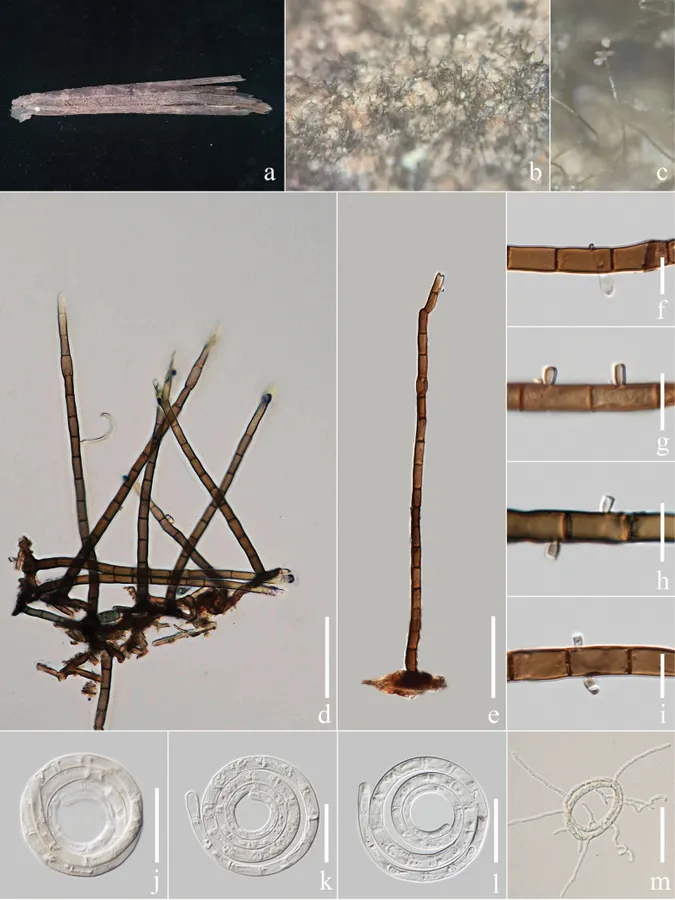
Helicoma rufum (MHZU 23-0168, new host record) a specimen observed b, c colony on decaying Caryota mitis d, e conidiophores f–i conidiogenous cells j–l conidia m germinated conidium. Scale bars: 50 μm (d, e, m); 10 μm (f–i); 20 μm (j–l).
- Jenis
- Pelat ilmiah
- Kredit
- Yinru Xiong, Kevin D. Hyde, Li Lu, Dulanjalee L. Harishchandra, Ausana Mapook, Biao Xu, Fatimah Alotibi, Ishara S. Manawasinghe
- Lisensi
- CC BY 4.0
- Sumber media
- Buka entri sumber
Sumber profil
- [1] Novel Helicoma and Neohelicosporium (Tubeufiaceae, Tubeufiales) species and two new host records of Helicoma on tropical palms (Arecaceae) from China — Helicoma rufum
- [2] Catalogue of Life Base2026-09-11 — Helicoma rufum
- [3] Dokumentasi visual — Helicoma rufum
Rujukan halaman ini
Fungi. Helicoma rufum. Rilis data fungi-2026-10-12-expansion-2. https://fungi-atlas.mgadib.chatgpt.site/jamur/helicoma-rufum/.
Status: impor sumber; pemeriksaan editorial per profil belum selesai. Untuk klaim biologis, gunakan rujukan pada bagian terkait dan periksa konteks sumber aslinya.