Ciri dan struktur
Subiculum annual, arachnoid, white, effused forming irregular areas to 5 x 2.5 cm. Pilei scattered or crowded in small groups, not confluent, cylindrical, 0.25-1.25 mm tall, 0.1-0.3 mm diameter, attached by narrow bases, ceraceous, fragile, white drying honey yellow; pileus surface delicately pruinose, clothed with delicate septate abhymenial hairs bearing apically 3-11 filiform branches 0.5-1 µm diameter; margin even, fringed with a compact palisade of branched hairs, erect or slightly inturned. Context white, to 30 µm thick, of densely compacted parallel hyphae; generative hyphae 2-2.5 µm diameter, walls 0.2 µm thick, hyaline. Hymenial layer to 25 µm deep, a close palisade of basidia and paraphyses. Basidia clavate, 14-18 x 4-5.5 µm, bearing 4 spores; sterigmata erect, slender, to 4 µm long. Paraphyses clavate, 8-14 x 3.5-4 µm. Spores globose or subglobose, apiculate, 4.5-5 µm diameter, walls smooth, hyaline, 0.2 µm thick.
Habitat dan peran ekologis
DISTRIBUTION: Europe, Great Britain, North America, New Zealand. HABITAT: Solitary or crowded on bark or decorticated wood of dead branches.
- Habitat atau konteks temuan
- Informasi belum tersedia pada sumber rilis ini.
- Substrat
- kulit kayu
- Peran atau konteks
- Belum dikategorikan pada rilis ini.
Reproduksi
Subiculum annual, arachnoid, white, effused forming irregular areas to 5 x 2.5 cm. Pilei scattered or crowded in small groups, not confluent, cylindrical, 0.25-1.25 mm tall, 0.1-0.3 mm diameter, attached by narrow bases, ceraceous, fragile, white drying honey yellow; pileus surface delicately pruinose, clothed with delicate septate abhymenial hairs bearing apically 3-11 filiform branches 0.5-1 µm diameter; margin even, fringed with a compact palisade of branched hairs, erect or slightly inturned. Context white, to 30 µm thick, of densely compacted parallel hyphae; generative hyphae 2-2.5 µm diameter, walls 0.2 µm thick, hyaline. Hymenial layer to 25 µm deep, a close palisade of basidia and paraphyses.
Catatan identitas dan cakupan
- Nama diterima mengikuti Catalogue of Life Base2026-09-11; uraian mempertahankan konsep dan nama sumber aslinya.
- Historical description as cited by BiotaNZ. Preserve explicit New Zealand/locality, host and specimen qualifiers. Distribution is the cited source scope, not a current global range.
- Impor dan terjemahan otomatis belum diperiksa editorial per profil. Struktur reproduktif yang disebutkan tidak membuktikan seluruh siklus hidup diketahui.
- Pengukuran pada uraian mengikuti spesimen, kultur, dan metode sumber; angka tidak diperluas ke semua populasi.
Catatan distribusi dalam narasi mengikuti sumbernya. Dokumentasi spesimen atau foto mengikuti catatan sumber. Bukti wilayah Indonesia dan penilaian konservasi tidak disimpulkan dari gambar.
Melihat dengan konteks
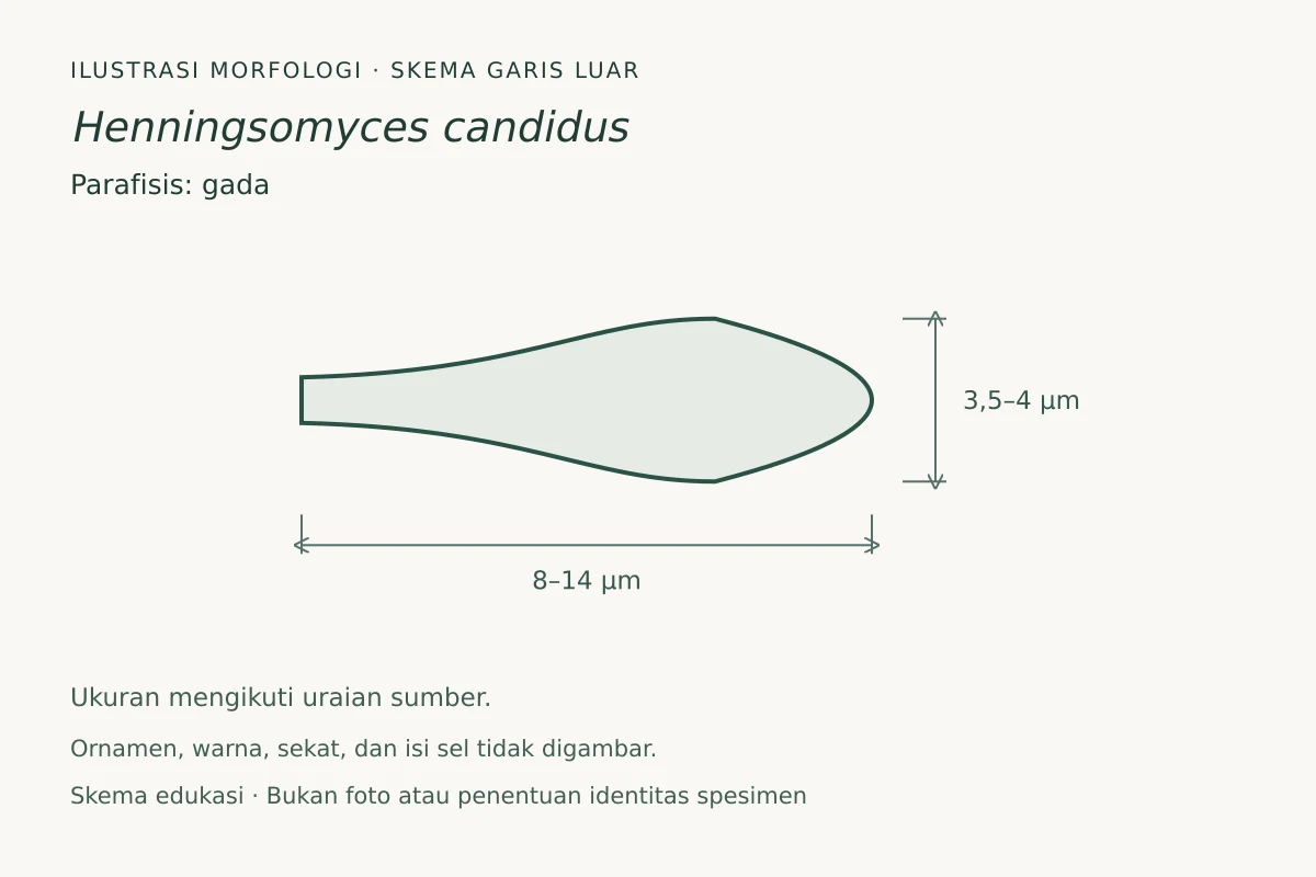
Ilustrasi skematis garis luar parafisis Henningsomyces candidus, berdasarkan uraian sumber; ukuran yang dilaporkan 8–14 × 3,5–4 µm. Proporsi disederhanakan, bukan salinan satu spesimen. Ornamen permukaan, warna, sekat, dan isi sel tidak direkonstruksi. Parafisis berbentuk gada, 8–14 × 3,5–4 µm. Rambut bercabang, sterigma basidium, dan apikulus spora merupakan struktur berbeda. Ornamen, warna, sekat, dan isi sel tidak digambar. Skema edukasi, bukan alat identifikasi.
- Jenis
- Ilustrasi skematis
- Kredit
- Fungi, ilustrasi orisinal (2026); fakta morfologi berdasarkan Cunningham, G.H. 1963: The Thelephoraceae of Australia and New Zealand. New Zealand Department of Scientific and Industrial Research, Bulletin. 145.
- Lisensi
- CC BY-SA 4.0
- Sumber media
- Buka entri sumber
Sumber profil
- [1] Cunningham, G.H. 1963: The Thelephoraceae of Australia and New Zealand. New Zealand Department of Scientific and Industrial Research, Bulletin. 145. — Lachnella candida
- [2] Catalogue of Life Base2026-09-11 — Henningsomyces candidus
Rujukan halaman ini
Fungi. Henningsomyces candidus. Rilis data fungi-2026-10-12-expansion-2. https://fungi-atlas.mgadib.chatgpt.site/jamur/henningsomyces-candidus/.
Status: impor sumber; pemeriksaan editorial per profil belum selesai. Untuk klaim biologis, gunakan rujukan pada bagian terkait dan periksa konteks sumber aslinya.