Ciri dan struktur
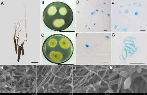
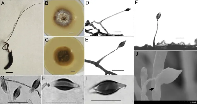
Hirsutella kuankuoshuiensis differs from other species in this genus primarily by its clavate, narrow fusiform or botuliform conidia and subulate or slender columnar phialide. Synnemata are single, extending from the head of insect; 8.6 cm long, dark brown and changing to brown towards the apex; no conidiation was observed (Fig. 3A). The fungus spreads slowly on PDA agar at 20–22 °C and grows to a diam. of 22–30 mm after 14 d; the colony is round, centre of surface with brown dense bulges and grey-white sparse flocculent aerial hyphae. Colony margin is flat with radial groove; a large amount of brown pigment secreted into the medium causes the back of colony to appear dark brown; thickness 10–12 mm (Fig. 3B, C). Mycelium hyaline, smooth, septate, 1.5–3.0 μm wide. Conidiogenous cells monophialidic, hyaline, borne perpendicular or at an acute angle to the subtending hyphae. Phialides subulate or slender columnar, tapering gradually to a long and narrow neck, 30–45 × 1–3 μm long. Conidia clavate, narrow fusiform or botuliform without a diaphragm, 9.9–12.6 × 2.7–4.5 μm, single- or double-enveloped in a hyaline mucus, thickness 2.0–3.0 μm (Fig. 3D–J).
Habitat dan peran ekologis
Synnemata are single, extending from the head of insect; 8.6 cm long, dark brown and changing to brown towards the apex; no conidiation was observed (Fig. 3A). The fungus spreads slowly on PDA agar at 20–22 °C and grows to a diam. of 22–30 mm after 14 d; the colony is round, centre of surface with brown dense bulges and grey-white sparse flocculent aerial hyphae. Colony margin is flat with radial groove; a large amount of brown pigment secreted into the medium causes the back of colony to appear dark brown; thickness 10–12 mm (Fig. 3B, C). Mycelium hyaline, smooth, septate, 1.5–3.0 μm wide. Conidiogenous cells monophialidic, hyaline, borne perpendicular or at an acute angle to the subtending hyphae. Phialides subulate or slender columnar, tapering gradually to a long and narrow neck, 30–45 × 1–3 μm long. Conidia clavate, narrow fusiform or botuliform without a diaphragm, 9.9–12.6 × 2.7–4.5 μm, single- or double-enveloped in a hyaline mucus, thickness 2.0–3.0 μm (Fig. 3D–J).
- Habitat atau konteks temuan
- kultur yang didokumentasikan
- Substrat
- Informasi belum tersedia pada sumber rilis ini.
- Peran atau konteks
- Belum dikategorikan pada rilis ini.
Reproduksi
Hirsutella kuankuoshuiensis differs from other species in this genus primarily by its clavate, narrow fusiform or botuliform conidia and subulate or slender columnar phialide. Synnemata are single, extending from the head of insect; 8.6 cm long, dark brown and changing to brown towards the apex; no conidiation was observed (Fig. 3A). The fungus spreads slowly on PDA agar at 20–22 °C and grows to a diam. of 22–30 mm after 14 d; the colony is round, centre of surface with brown dense bulges and grey-white sparse flocculent aerial hyphae. Colony margin is flat with radial groove; a large amount of brown pigment secreted into the medium causes the back of colony to appear dark brown; thickness 10–12 mm (Fig. 3B, C).
Catatan identitas dan cakupan
- Nama diterima mengikuti Catalogue of Life Base2026-09-11; uraian mempertahankan konsep dan nama sumber aslinya.
- Preserve original treatment localities, hosts and substrate scope. Materials examined describe documented specimens, not the full global range.
- Impor dan terjemahan otomatis belum diperiksa editorial per profil. Struktur reproduktif yang disebutkan tidak membuktikan seluruh siklus hidup diketahui.
- Pengukuran pada uraian mengikuti spesimen, kultur, dan metode sumber; angka tidak diperluas ke semua populasi.
Catatan distribusi dalam narasi mengikuti sumbernya. Dokumentasi spesimen atau foto mengikuti catatan sumber. Bukti wilayah Indonesia dan penilaian konservasi tidak disimpulkan dari gambar.
Melihat dengan konteks
Morphological characteristics of Hirsutella kuankuoshuiensis A the insect specimens with single and thin synnemata (HKAS112885) B, C colonial morphology on PDA agar media for 20 d B shows the front of the colony and C shows the back of the colony D–G LM images showing conidiogenous cells and conidia D, E the structure of conidiogenous cells on mycelia F the images of conidiogenous cells on synnemata (optical microscope) H–J conidial morphology (LM) G conidia with mucilage (SEM). Scale bars: 10 mm (A–C); bar of G was shown in the figure; the rest of the bars were 10 μm. LM, light microscopy; PDA, potato dextrose agar; SEM, scanning electron microscopy.
- Jenis
- Pelat ilmiah
- Kredit
- Jiaojiao Qu, Xiao Zou, Wei Cao, Zhongshun Xu, Zongqi Liang
- Lisensi
- CC BY 4.0
- Sumber media
- Buka entri sumber
Sumber profil
- [1] Two new species of Hirsutella (Ophiocordycipitaceae, Sordariomycetes) that are parasitic on lepidopteran insects from China — Hirsutella kuankuoshuiensis
- [2] Catalogue of Life Base2026-09-11 — Hirsutella kuankuoshuiensis
- [3] Dokumentasi visual — Hirsutella kuankuoshuiensis
Rujukan halaman ini
Fungi. Hirsutella kuankuoshuiensis. Rilis data fungi-2026-10-12-expansion-2. https://fungi-atlas.mgadib.chatgpt.site/jamur/hirsutella-kuankuoshuiensis/.
Status: impor sumber; pemeriksaan editorial per profil belum selesai. Untuk klaim biologis, gunakan rujukan pada bagian terkait dan periksa konteks sumber aslinya.