Ciri dan struktur
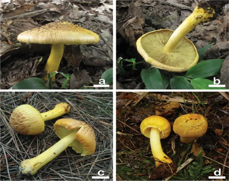
Small to medium-sized. Pileus: 30–60 mm in diameter, bell-shaped to convex when young, and then planar with umbonate center; margin strongly in-rolled or deflexed when young, and then gradually straight when mature; center covered with stout, erect, conic squamules (up to 2 mm high, 1–1.5 mm wide), coarsely fibrillose towards the margin; surface dry, primrose yellow (1A6) to bright yellow (2A5), becoming pale brown (3B6) over the disc. Lamellae crowded (ca. 50–70), 3–5 mm wide, adnexed to adnato-decurrent, often subsinuate; light yellow (1A5) turning to pale yellow-fuscous (2B5), edge concolorous, even. Stipe 35–75 × 4–8 mm, cylindrical or attenuated towards apex, stout, base subbulbous to bulbous, up to 16 mm wide; bright yellow (2A5); apex pruinose, covered with bright yellow(2A5) to orange (2A6), longitudinal, floccose-fibrillose fibrils towards base; dry, solid. Cortina conspicuous present in young specimens. Context pale yellow (1A4) in stipe and cuticle. (5.0) 5.5–9.0 (10.0) µm (av. 7.1 μm, SD 1.1 μm) × (4.0) 4.5–6.0 (6.5) µm (av. 5.3 μm, SD 0.6 μm), Q = (1.00) 1.11–1.67 (1.80), Qm = 1.33 ± 0.19 (n = 200 of 10 coll.), nodulose, 6–8 hemispheric knobs, yellow-brown with 5% KOH. Basidia: 17–26 × 7–9 µm, 4-spored, clavate to broadly clavate. Pleurocystidia: 37–67 µm (av. 46.1 μm, SD 3.0 μm) × 10–18 µm (av. 13.4 μm, SD 1.2 μm), Q = 2.80–4.0 (n = 100 of 10 coll.), abundant, broadly fusoid to lageniform; crystalliferous at apex, base usually truncate to obtuse, occasionally tapered into pedicel; metuloid, hyaline, sometimes contain a few small crystals or resinous inclusions, thick-walled, walls up to 1.5 µm thick, bright yellow with 5% KOH.
Habitat dan peran ekologis
Single to scattered in mixed forest dominated by Pinus and Quercus. Malaysia (type location) (Horak 1979), China (Hunan Province, Anhui Province).
- Habitat atau konteks temuan
- hutan
- Substrat
- Informasi belum tersedia pada sumber rilis ini.
- Peran atau konteks
- Belum dikategorikan pada rilis ini.
Reproduksi
(5.0) 5.5–9.0 (10.0) µm (av. 7.1 μm, SD 1.1 μm) × (4.0) 4.5–6.0 (6.5) µm (av. 5.3 μm, SD 0.6 μm), Q = (1.00) 1.11–1.67 (1.80), Qm = 1.33 ± 0.19 (n = 200 of 10 coll.), nodulose, 6–8 hemispheric knobs, yellow-brown with 5% KOH. Basidia: 17–26 × 7–9 µm, 4-spored, clavate to broadly clavate. Pleurocystidia: 37–67 µm (av. 46.1 μm, SD 3.0 μm) × 10–18 µm (av. 13.4 μm, SD 1.2 μm), Q = 2.80–4.0 (n = 100 of 10 coll.), abundant, broadly fusoid to lageniform; crystalliferous at apex, base usually truncate to obtuse, occasionally tapered into pedicel; metuloid, hyaline, sometimes contain a few small crystals or resinous inclusions, thick-walled, walls up to 1.5 µm thick, bright yellow with 5% KOH. Cheilocystidia similar to pleurocystidia, 35–62 × 9–17 µm; paracystidia: 12–25 × 5–11 µm, abundant, thin-walled, translucent inside, clavate to broadly clavate.
Catatan identitas dan cakupan
- Nama diterima mengikuti Catalogue of Life Base2026-09-11; uraian mempertahankan konsep dan nama sumber aslinya.
- Preserve original treatment localities, hosts and substrate scope. Materials examined describe documented specimens, not the full global range.
- Impor dan terjemahan otomatis belum diperiksa editorial per profil. Struktur reproduktif yang disebutkan tidak membuktikan seluruh siklus hidup diketahui.
- Pengukuran pada uraian mengikuti spesimen, kultur, dan metode sumber; angka tidak diperluas ke semua populasi.
Catatan distribusi dalam narasi mengikuti sumbernya. Dokumentasi spesimen atau foto mengikuti catatan sumber. Bukti wilayah Indonesia dan penilaian konservasi tidak disimpulkan dari gambar.
Melihat dengan konteks
Basidiomata of Inocybe squarrosolutea a, b MHHNU8536 c MHHNU31006, and d MHHNU31427. Scale bars: 10 mm.
- Jenis
- Pelat ilmiah
- Kredit
- Sai Nan Li, Fei Xu, Ming Jiang, Feng Liu, Fang Wu, Ping Zhang, Yu Guang Fan, Zuo Hong Chen
- Lisensi
- CC BY 4.0
- Sumber media
- Buka entri sumber
Dokumentasi tambahan
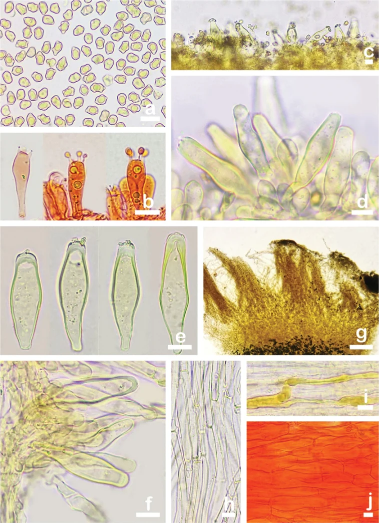
Microscopic features of Inocybe squarrosolutea (MHHNU31427) a basidiospores b basidia with probasidium c gill edge d cheilocystidia and paracystidia e pleurocystidia f caulocystidia and cauloparacystidia g, h pileipellis i oleiferous hyphae, and j hymenial hyphae. Scale bars: 10 µm.
Kredit dan penggunaan
Sai Nan Li, Fei Xu, Ming Jiang, Feng Liu, Fang Wu, Ping Zhang, Yu Guang Fan, Zuo Hong Chen
Ukuran dan format gambar disesuaikan tanpa pemotongan, retus, atau penambahan skala. Lisensi sumber dipertahankan.
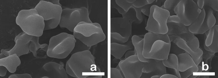
Pelat perbandingan basidiospora pada SEM. Inocybe squarrosolutea ditampilkan pada panel a; panel lainnya memuat spesies pembanding. Pembagian panel mengikuti keterangan sumber.
Kredit dan penggunaan
Sai Nan Li, Fei Xu, Ming Jiang, Feng Liu, Fang Wu, Ping Zhang, Yu Guang Fan, Zuo Hong Chen
Ukuran dan format gambar disesuaikan tanpa pemotongan, retus, atau penambahan skala. Lisensi sumber dipertahankan.
Sumber profil
- [1] Two new toxic yellow Inocybe species from China: morphological characteristics, phylogenetic analyses and toxin detection — Inocybe squarrosolutea
- [2] Catalogue of Life Base2026-09-11 — Inocybe squarrosolutea
- [3] Dokumentasi visual — Inocybe squarrosolutea
- [4] Dokumentasi visual — Inocybe squarrosolutea
- [5] Dokumentasi visual — Inocybe squarrosolutea
Rujukan halaman ini
Fungi. Inocybe squarrosolutea. Rilis data fungi-2026-10-12-expansion-2. https://fungi-atlas.mgadib.chatgpt.site/jamur/inocybe-squarrosolutea/.
Status: impor sumber; pemeriksaan editorial per profil belum selesai. Untuk klaim biologis, gunakan rujukan pada bagian terkait dan periksa konteks sumber aslinya.