01 / Morfologi

Ciri dan struktur

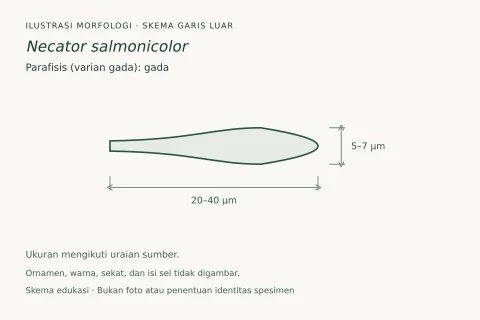
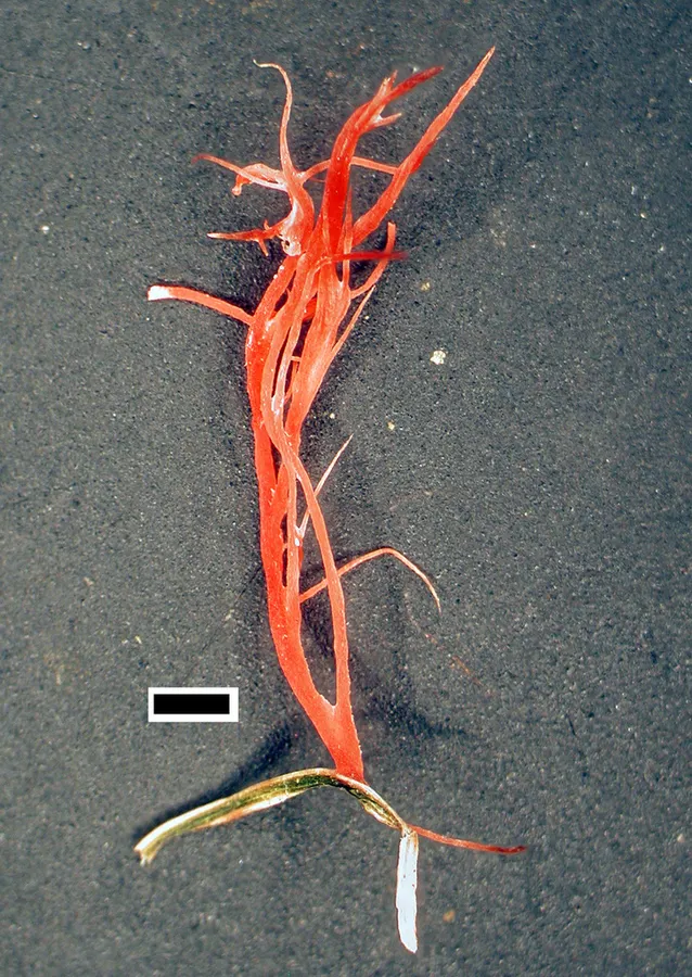
Hymenophore annual, ceraceous, growing from surfaces of culms and leaves of grasses, as clavariform or fuciform single or branched sometimes antler-like processes, each long-aculeate or subulate, 5-25 mm long; hymenial surface flesh-pink or coral-pink, remaining so or drying pallid tan. Context of closely compacted parallel hyphae often breaking into oidia near apices, intermediate layer scanty or wanting; generative hyphae 6-7 µm diameter, walls 0.2 µm, thick, naked or encrusted with orange mucilage granules, without clamp connections. Hymenial layer developing both upon the exterior of host tissues and the subulate processes, a dense palisade of basidia and paraphyses. Basidia subclavate, 16-20 x 5-6 µm, bearing 4 spores; sterigmata stout, to 6 µm long. Paraphyses subclavate, a few slightly capitate, 16-18 x 5-6 µm. Spores elliptical, with rounded ends, a few irregularly suballantoid, apiculate, 8-11 x 4-5 µm, walls smooth, hyaline, 0.2 µm thick.

Sebagian uraian ditampilkan dalam bahasa sumber; terjemahannya belum tersedia atau belum lolos pemeriksaan.

Rujukan: [1]

Pengukuran mengikuti objek, spesimen atau kultur, dan metode yang disebutkan sumber. Baca uraian asli untuk konteks lengkap.

02 / Kehidupan

Habitat dan peran ekologis

DISTRIBUTION: Great Britain, North America, Australia, Tasmania, New Zealand. HABITAT: Parasitic upon culms and leaves of grasses.

Sebagian uraian ditampilkan dalam bahasa sumber; terjemahannya belum tersedia atau belum lolos pemeriksaan.

Rujukan: [1]

Habitat atau konteks temuan
Informasi belum tersedia pada sumber rilis ini.
Substrat
Informasi belum tersedia pada sumber rilis ini.
Peran atau konteks
Belum dikategorikan pada rilis ini.
Temukan hubungan serupa
03 / Siklus hidup

Reproduksi

Hymenophore annual, ceraceous, growing from surfaces of culms and leaves of grasses, as clavariform or fuciform single or branched sometimes antler-like processes, each long-aculeate or subulate, 5-25 mm long; hymenial surface flesh-pink or coral-pink, remaining so or drying pallid tan. Context of closely compacted parallel hyphae often breaking into oidia near apices, intermediate layer scanty or wanting; generative hyphae 6-7 µm diameter, walls 0.2 µm, thick, naked or encrusted with orange mucilage granules, without clamp connections. Hymenial layer developing both upon the exterior of host tissues and the subulate processes, a dense palisade of basidia and paraphyses. Basidia subclavate, 16-20 x 5-6 µm, bearing 4 spores; sterigmata stout, to 6 µm long.

Sebagian uraian ditampilkan dalam bahasa sumber; terjemahannya belum tersedia atau belum lolos pemeriksaan.

Rujukan: [1]

Sinonim menurut acuan taksonomi rilis: Athelia fuciformis; Corticium fuciforme; Epithele fuciformis; Hypochnus fuciformis; Isaria fuciformis; Isaria graminiperda var. fuciformis; Phanerochaete fuciformis; Xylaria fuciformis
04 / Batas informasi

Catatan identitas dan cakupan

  • Nama diterima mengikuti Catalogue of Life Base2026-09-11; uraian mempertahankan konsep dan nama sumber aslinya.
  • Historical description as cited by BiotaNZ. Preserve explicit New Zealand/locality, host and specimen qualifiers. Distribution is the cited source scope, not a current global range.
  • Impor dan terjemahan otomatis belum diperiksa editorial per profil. Struktur reproduktif yang disebutkan tidak membuktikan seluruh siklus hidup diketahui.
  • Pengukuran pada uraian mengikuti spesimen, kultur, dan metode sumber; angka tidak diperluas ke semua populasi.

Catatan distribusi dalam narasi mengikuti sumbernya. Dokumentasi spesimen atau foto mengikuti catatan sumber. Bukti wilayah Indonesia dan penilaian konservasi tidak disimpulkan dari gambar.

05 / Dokumentasi

Melihat dengan konteks

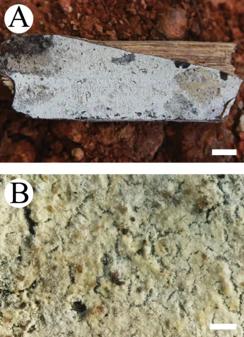
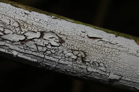
Dokumentasi Laetisaria fuciformis menurut asosiasi foto pada record takson iNaturalist. Kredit: Jerry Cooper. Identitas mengikuti sumber; bukan penentuan spesimen independen.

Jenis
Fotografi
Kredit
(c) Jerry Cooper, some rights reserved (CC BY), uploaded by Jerry Cooper
Lisensi
CC BY 4.0
Sumber media
Buka entri sumber

Gambar menunjukkan objek menurut identifikasi sumber, bukan verifikasi independen spesimen oleh Fungi. Tidak ada skala baru yang ditambahkan.

06 / Rujukan

Sumber profil

  1. [1] Cunningham, G.H. 1963: The Thelephoraceae of Australia and New Zealand. New Zealand Department of Scientific and Industrial Research, Bulletin. 145. — Corticium fuciformeBagian sumber: biota:1cb0defb-36b9-11d5-9548-00d0592d548c:1cb1bae8-36b9-11d5-9548-00d0592d548c
    Tanggal penghimpunan sumber: 2026-10-12
    Teks sumber: CC BY 4.0 · Cunningham, G.H. 1963: The Thelephoraceae of Australia and New Zealand. New Zealand Department of Scientific and Industrial Research, Bulletin. 145.
  2. [2] Catalogue of Life Base2026-09-11 — Laetisaria fuciformisBagian sumber: ColDP NameUsage.tsv ID 3RV84; accepted species; source 2073
    Tanggal penghimpunan sumber: 2026-10-12
  3. [3] Dokumentasi visual — Laetisaria fuciformisBagian sumber: Individual media record; identification follows the source, not independent determination
    Tanggal penghimpunan sumber: 2026-10-12
    Media sumber: CC BY 4.0 · (c) Jerry Cooper, some rights reserved (CC BY), uploaded by Jerry Cooper

Adaptasi teks: CC BY 4.0. Cunningham, G.H. 1963: The Thelephoraceae of Australia and New Zealand. New Zealand Department of Scientific and Industrial Research, Bulletin. 145. — Cunningham, G.H. 1963: The Thelephoraceae of Australia and New Zealand. New Zealand Department of Scientific and Industrial Research, Bulletin. 145. — Corticium fuciforme

Uraian asli dan riwayat sumber

Riwayat atau halaman sumber

Ringkasan sumber

Hymenophore annual, ceraceous, growing from surfaces of culms and leaves of grasses, as clavariform or fuciform single or branched sometimes antler-like processes, each long-aculeate or subulate, 5-25 mm long; hymenial surface flesh-pink or coral-pink, remaining so or drying pallid tan. Context of closely compacted parallel hyphae often breaking into oidia near apices, intermediate layer scanty or wanting; generative hyphae 6-7 µm diameter, walls 0.2 µm, thick, naked or encrusted with orange mucilage granules, without clamp connections.

Revisi sumber

Morfologi sumber

Hymenophore annual, ceraceous, growing from surfaces of culms and leaves of grasses, as clavariform or fuciform single or branched sometimes antler-like processes, each long-aculeate or subulate, 5-25 mm long; hymenial surface flesh-pink or coral-pink, remaining so or drying pallid tan. Context of closely compacted parallel hyphae often breaking into oidia near apices, intermediate layer scanty or wanting; generative hyphae 6-7 µm diameter, walls 0.2 µm, thick, naked or encrusted with orange mucilage granules, without clamp connections. Hymenial layer developing both upon the exterior of host tissues and the subulate processes, a dense palisade of basidia and paraphyses. Basidia subclavate, 16-20 x 5-6 µm, bearing 4 spores; sterigmata stout, to 6 µm long. Paraphyses subclavate, a few slightly capitate, 16-18 x 5-6 µm. Spores elliptical, with rounded ends, a few irregularly suballantoid, apiculate, 8-11 x 4-5 µm, walls smooth, hyaline, 0.2 µm thick.

Revisi sumber

Ekologi sumber

DISTRIBUTION: Great Britain, North America, Australia, Tasmania, New Zealand. HABITAT: Parasitic upon culms and leaves of grasses.

Revisi sumber

Struktur reproduksi sumber

Hymenophore annual, ceraceous, growing from surfaces of culms and leaves of grasses, as clavariform or fuciform single or branched sometimes antler-like processes, each long-aculeate or subulate, 5-25 mm long; hymenial surface flesh-pink or coral-pink, remaining so or drying pallid tan. Context of closely compacted parallel hyphae often breaking into oidia near apices, intermediate layer scanty or wanting; generative hyphae 6-7 µm diameter, walls 0.2 µm, thick, naked or encrusted with orange mucilage granules, without clamp connections. Hymenial layer developing both upon the exterior of host tissues and the subulate processes, a dense palisade of basidia and paraphyses. Basidia subclavate, 16-20 x 5-6 µm, bearing 4 spores; sterigmata stout, to 6 µm long.

Revisi sumber

Rujukan halaman ini

Fungi. Laetisaria fuciformis. Rilis data fungi-2026-10-12-expansion-2. https://fungi-atlas.mgadib.chatgpt.site/jamur/laetisaria-fuciformis/.

Status: impor sumber; pemeriksaan editorial per profil belum selesai. Untuk klaim biologis, gunakan rujukan pada bagian terkait dan periksa konteks sumber aslinya.