Saprobic on cabang mati dari Urtica dioica. Ascomata 100–130 tinggi, 70–110 μm berlian, sepi, tersebar atau dalam kelompok kecil, beres melalui epidermis inang menjadi superficial, konik menjadi mammiform, cokelat gelap menjadi hitam, coriaceous, halus, mudah dikeluarkan dari substrat inang, ostiolate dengan papilla kecil. Osteoles 50–70 μm berlian, papillate, hitam, berkilau, halus. Peridium 25–50 μm wide, comprising two cell types, outer layer composed of small, thick-walled cells of textura angularis to textura globulosa, surface heavily pigmented termed as scleroplectenchyma, thinner at the apex, wide at sides, inner layer composed of subhyaline or light brown relatively thin-walled cells of textura angularis, cells near the base comparatively larger.
Sebagian uraian ditampilkan dalam bahasa sumber; terjemahannya belum tersedia atau belum lolos pemeriksaan.
Profil ini diimpor dari uraian spesies pada sumber berlisensi dan sebagian teks diterjemahkan otomatis. Pemeriksaan editorial per profil belum selesai. Bagian yang belum diterjemahkan atau belum lolos pemeriksaan terjemahan tetap ditampilkan dalam bahasa sumber. Angka dan nama ilmiah dipertahankan.
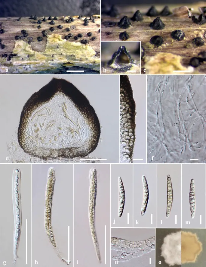
Saprobic on dead branches of Urtica dioica. Sexual morph Ascomata 100–130 high, 70–110 μm diam., solitary, scattered or in small groups, erumpent through host epidermis to superficial, conical to mammiform, dark brown to black, coriaceous, smooth, easily removed from the host substrate, ostiolate with minute papilla. Ostioles 50–70 μm diam., papillate, black, shiny, smooth. Peridium 25–50 μm wide, comprising two cell types, outer layer composed of small, thick-walled cells of textura angularis to textura globulosa, surface heavily pigmented termed as scleroplectenchyma, thinner at the apex, wide at sides, inner layer composed of subhyaline or light brown relatively thin-walled cells of textura angularis, cells near the base comparatively larger. Hamathecium comprising numerous, dense, 1.5–2 μm wide, filamentous, septate, cellular pseudoparaphyses, branched and anastomosing, embedded in gelatinous matrix. Asci 60–140 × 9–11 μm (\(\bar{x} \) = 104.5 × 10 μm, n = 20), 8-spored, bitunicate, fissitunicate, cylindrical, rounded at the apex, pedicellate, numerous, with ocular chamber. Ascospores 35–40 × 4–6 μm (\(\bar{x} \) = 38.3 × 5.2 μm, n = 20), overlapping 1–2-seriate, initially hyaline, becoming yellowish brown at maturity, long fusiform, (8–)9-septate, constricted at the septa, narrowly rounded at both ends, smooth-walled, lacking a mucilaginous sheath. Asexual morph Undetermined. Culture characteristics: Ascospores germinating on MEA within 24 h. Colonies growing on MEA reaching 2 cm diam. in one week at 16 °C. Mycelium superficial, surface smooth, irregular, slightly raised, edge crenate, velutinous, from above white to pale yellow, reverse yellow.
Sebagian uraian ditampilkan dalam bahasa sumber; terjemahannya belum tersedia atau belum lolos pemeriksaan.
Pengukuran mengikuti objek, spesimen atau kultur, dan metode yang disebutkan sumber. Baca uraian asli untuk konteks lengkap.
02 / Kehidupan
Habitat dan peran ekologis
Saprobic on dead branches of Urtica dioica. Sexual morph Ascomata 100–130 high, 70–110 μm diam., solitary, scattered or in small groups, erumpent through host epidermis to superficial, conical to mammiform, dark brown to black, coriaceous, smooth, easily removed from the host substrate, ostiolate with minute papilla. Ostioles 50–70 μm diam., papillate, black, shiny, smooth. Peridium 25–50 μm wide, comprising two cell types, outer layer composed of small, thick-walled cells of textura angularis to textura globulosa, surface heavily pigmented termed as scleroplectenchyma, thinner at the apex, wide at sides, inner layer composed of subhyaline or light brown relatively thin-walled cells of textura angularis, cells near the base comparatively larger. Hamathecium comprising numerous, dense, 1.5–2 μm wide, filamentous, septate, cellular pseudoparaphyses, branched and anastomosing, embedded in gelatinous matrix. Asci 60–140 × 9–11 μm (\(\bar{x} \) = 104.5 × 10 μm, n = 20), 8-spored, bitunicate, fissitunicate, cylindrical, rounded at the apex, pedicellate, numerous, with ocular chamber. Ascospores 35–40 × 4–6 μm (\(\bar{x} \) = 38.3 × 5.2 μm, n = 20), overlapping 1–2-seriate, initially hyaline, becoming yellowish brown at maturity, long fusiform, (8–)9-septate, constricted at the septa, narrowly rounded at both ends, smooth-walled, lacking a mucilaginous sheath.
Sebagian uraian ditampilkan dalam bahasa sumber; terjemahannya belum tersedia atau belum lolos pemeriksaan.
Saprobic on dead branches of Urtica dioica. Sexual morph Ascomata 100–130 high, 70–110 μm diam., solitary, scattered or in small groups, erumpent through host epidermis to superficial, conical to mammiform, dark brown to black, coriaceous, smooth, easily removed from the host substrate, ostiolate with minute papilla. Ostioles 50–70 μm diam., papillate, black, shiny, smooth. Peridium 25–50 μm wide, comprising two cell types, outer layer composed of small, thick-walled cells of textura angularis to textura globulosa, surface heavily pigmented termed as scleroplectenchyma, thinner at the apex, wide at sides, inner layer composed of subhyaline or light brown relatively thin-walled cells of textura angularis, cells near the base comparatively larger.
Sebagian uraian ditampilkan dalam bahasa sumber; terjemahannya belum tersedia atau belum lolos pemeriksaan.
Nama diterima mengikuti Catalogue of Life Base2026-09-11; uraian mempertahankan konsep dan nama sumber aslinya.
Preserve original monograph treatment localities, hosts and specimen/substrate scope. Materials examined document specimens, not the full global range.
Impor dan terjemahan otomatis belum diperiksa editorial per profil. Struktur reproduktif yang disebutkan tidak membuktikan seluruh siklus hidup diketahui.
Pengukuran pada uraian mengikuti spesimen, kultur, dan metode sumber; angka tidak diperluas ke semua populasi.
Catatan distribusi dalam narasi mengikuti sumbernya. Dokumentasi spesimen atau foto mengikuti catatan sumber. Bukti wilayah Indonesia dan penilaian konservasi tidak disimpulkan dari gambar.
05 / Dokumentasi
Melihat dengan konteks
Leptosphaeria urticae (MFLU 18-0591, holotype). a–c Appearance of ascomata on host surface. d Section through an ascoma. e Peridium. f Pseudoparaphyses. g–i Asci. j–m Ascospores. n Spore germination on MEA after 24 h. o Culture from above and below. Scale bars a, b = 500 µm, c = 200 μm, d = 100 μm, e, g–i = 50 μm, j–m = 10 μm, f = 5 μm
Jenis
Pelat ilmiah
Kredit
Penulis dan sumber artikel: Phookamsak, Rungtiwa; Hyde, Kevin D.; Jeewon, Rajesh; Bhat, D. Jayarama; Jones, E. B. Gareth; Maharachchikumbura, Sajeewa S. N.; Raspé, Olivier; Karunarathna, Samantha C.; Wanasinghe, Dhanushka N.; Hongsanan, Sinang; Doilom, Mingkwan; Tennakoon, Danushka S.; Machado, Alexandre R.; Firmino, André L.; Ghosh, Aniket; Karunarathna, Anuruddha; Mešić, Armin; Dutta, Arun Kumar; Thongbai, Benjarong; Devadatha, Bandarupalli; Norphanphoun, Chada; Senwanna, Chanokned; Wei, Deping; Pem, Dhandevi; Ackah, Frank Kwekucher; Wang, Gen-Nuo; Jiang, Hong-Bo; Madrid, Hugo; Lee, Hyang Burm; Goonasekara, Ishani D.; Manawasinghe, Ishara S.; Kušan, Ivana; Cano, Josep; Gené, Josepa; Li, Junfu; Das, Kanad; Acharya, Krishnendu; Raj, K. N. Anil; Latha, K. P. Deepna; Chethana, K. W. Thilini; He, Mao-Qiang; Dueñas, Margarita; Jadan, Margita; Martín, María P.; Samarakoon, Milan C.; Dayarathne, Monika C.; Raza, Mubashar; Park, Myung Soo; Telleria, M. Teresa; Chaiwan, Napalai; Matočec, Neven; de Silva, Nimali I.; Pereira, Olinto L.; Singh, Paras Nath; Manimohan, Patinjareveettil; Uniyal, Priyanka; Shang, Qiu-Ju; Bhatt, Rajendra P.; Perera, Rekhani H.; Alvarenga, Renato Lúcio Mendes; Nogal-Prata, Sandra; Singh, Sanjay K.; Vadthanarat, Santhiti; Oh, Seung-Yoon; Huang, Shi-Ke; Rana, Shiwali; Konta, Sirinapa; Paloi, Soumitra; Jayasiri, Subashini C.; Jeon, Sun Jeong; Mehmood, Tahir; Gibertoni, Tatiana Baptista; Nguyen, Thuong T. T.; Singh, Upendra; Thiyagaraja, Vinodhini; Sarma, V. Venkateswara; Dong, Wei; Yu, Xian-Dong; Lu, Yong-Zhong; Lim, Young Woon; Chen, Yun; Tkalčec, Zdenko; Zhang, Zhi-Feng; Luo, Zong-Long; Daranagama, Dinushani A.; Thambugala, Kasun M.; Tibpromma, Saowaluck; Camporesi, Erio; Bulgakov, Timur S.; Dissanayake, Asha J.; Senanayake, Indunil C.; Dai, Dong Qin; Tang, Li-Zhou; Khan, Sehroon; Zhang, Huang; Promputtha, Itthayakorn; Cai, Lei; Chomnunti, Putarak; Zhao, Rui-Lin; Lumyong, Saisamorn; Boonmee, Saranyaphat; Wen, Ting-Chi; Mortimer, Peter E.; Xu, Jianchu
Saprobic on dead branches of Urtica dioica.
Sexual morph Ascomata 100–130 high, 70–110 μm diam., solitary, scattered or in small groups, erumpent through host epidermis to superficial, conical to mammiform, dark brown to black, coriaceous, smooth, easily removed from the host substrate, ostiolate with minute papilla.
Ostioles 50–70 μm diam., papillate, black, shiny, smooth.
Peridium 25–50 μm wide, comprising two cell types, outer layer composed of small, thick-walled cells of textura angularis to textura globulosa, surface heavily pigmented termed as scleroplectenchyma, thinner at the apex, wide at sides, inner layer composed of subhyaline or light brown relatively thin-walled cells of textura angularis, cells near the base comparatively larger.
Saprobic on dead branches of Urtica dioica.
Sexual morph Ascomata 100–130 high, 70–110 μm diam., solitary, scattered or in small groups, erumpent through host epidermis to superficial, conical to mammiform, dark brown to black, coriaceous, smooth, easily removed from the host substrate, ostiolate with minute papilla.
Ostioles 50–70 μm diam., papillate, black, shiny, smooth.
Peridium 25–50 μm wide, comprising two cell types, outer layer composed of small, thick-walled cells of textura angularis to textura globulosa, surface heavily pigmented termed as scleroplectenchyma, thinner at the apex, wide at sides, inner layer composed of subhyaline or light brown relatively thin-walled cells of textura angularis, cells near the base comparatively larger.
Hamathecium comprising numerous, dense, 1.5–2 μm wide, filamentous, septate, cellular pseudoparaphyses, branched and anastomosing, embedded in gelatinous matrix.
Asci 60–140 × 9–11 μm (\(\bar{x} \) = 104.5 × 10 μm, n = 20), 8-spored, bitunicate, fissitunicate, cylindrical, rounded at the apex, pedicellate, numerous, with ocular chamber.
Ascospores 35–40 × 4–6 μm (\(\bar{x} \) = 38.3 × 5.2 μm, n = 20), overlapping 1–2-seriate, initially hyaline, becoming yellowish brown at maturity, long fusiform, (8–)9-septate, constricted at the septa, narrowly rounded at both ends, smooth-walled, lacking a mucilaginous sheath.
Asexual morph Undetermined.
Culture characteristics: Ascospores germinating on MEA within 24 h.
Colonies growing on MEA reaching 2 cm diam. in one week at 16 °C.
Mycelium superficial, surface smooth, irregular, slightly raised, edge crenate, velutinous, from above white to pale yellow, reverse yellow.
Saprobic on dead branches of Urtica dioica.
Sexual morph Ascomata 100–130 high, 70–110 μm diam., solitary, scattered or in small groups, erumpent through host epidermis to superficial, conical to mammiform, dark brown to black, coriaceous, smooth, easily removed from the host substrate, ostiolate with minute papilla.
Ostioles 50–70 μm diam., papillate, black, shiny, smooth.
Peridium 25–50 μm wide, comprising two cell types, outer layer composed of small, thick-walled cells of textura angularis to textura globulosa, surface heavily pigmented termed as scleroplectenchyma, thinner at the apex, wide at sides, inner layer composed of subhyaline or light brown relatively thin-walled cells of textura angularis, cells near the base comparatively larger.
Hamathecium comprising numerous, dense, 1.5–2 μm wide, filamentous, septate, cellular pseudoparaphyses, branched and anastomosing, embedded in gelatinous matrix.
Asci 60–140 × 9–11 μm (\(\bar{x} \) = 104.5 × 10 μm, n = 20), 8-spored, bitunicate, fissitunicate, cylindrical, rounded at the apex, pedicellate, numerous, with ocular chamber.
Ascospores 35–40 × 4–6 μm (\(\bar{x} \) = 38.3 × 5.2 μm, n = 20), overlapping 1–2-seriate, initially hyaline, becoming yellowish brown at maturity, long fusiform, (8–)9-septate, constricted at the septa, narrowly rounded at both ends, smooth-walled, lacking a mucilaginous sheath.
Saprobic on dead branches of Urtica dioica.
Sexual morph Ascomata 100–130 high, 70–110 μm diam., solitary, scattered or in small groups, erumpent through host epidermis to superficial, conical to mammiform, dark brown to black, coriaceous, smooth, easily removed from the host substrate, ostiolate with minute papilla.
Ostioles 50–70 μm diam., papillate, black, shiny, smooth.
Peridium 25–50 μm wide, comprising two cell types, outer layer composed of small, thick-walled cells of textura angularis to textura globulosa, surface heavily pigmented termed as scleroplectenchyma, thinner at the apex, wide at sides, inner layer composed of subhyaline or light brown relatively thin-walled cells of textura angularis, cells near the base comparatively larger.
Status: impor sumber; pemeriksaan editorial per profil belum selesai. Untuk klaim biologis, gunakan rujukan pada bagian terkait dan periksa konteks sumber aslinya.
Lichtheimia corymbifera was originally described as Mucor corymbifer in 1884 by Lichtheim from clinical isolations in Wrocław, Poland. Spesies ini ditandai dengan columella berbentuk kerucut dan proyeksi pendek, jelas, apophysis berbentuk…