Ciri dan struktur
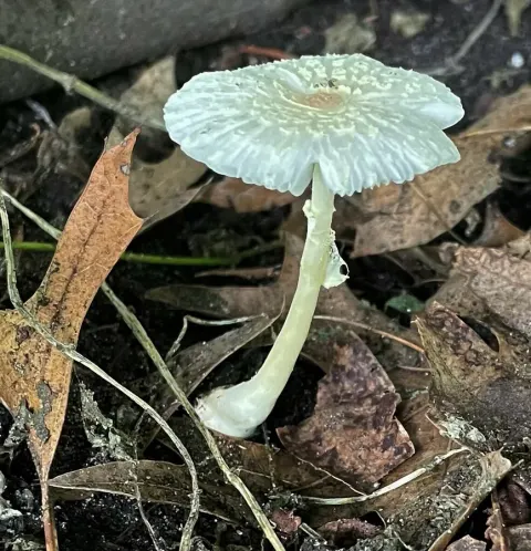
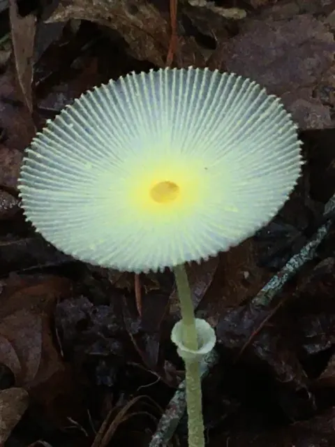
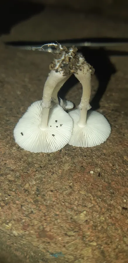
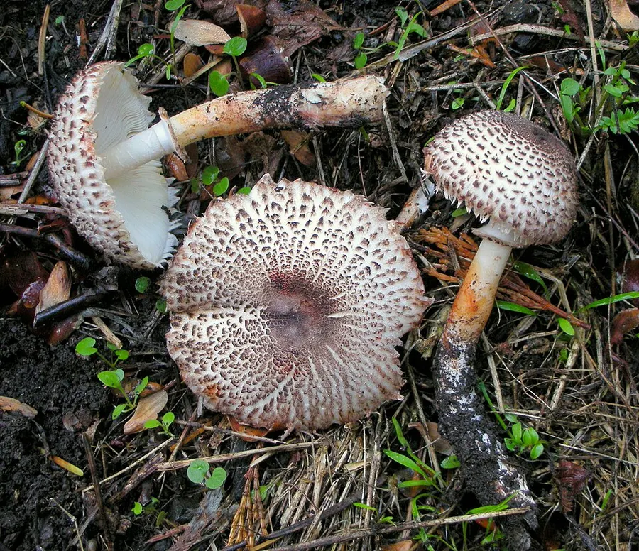
Its cap is 18-60 mm across. Its shape is initially hemispherical, then expanding to be convex or conical, ending up flat, or even slightly concave. It has dark brown scales arranged circularly around the purple to red to brown centre. When touched, the cap turns red-orange, which fades to dark-brown. The gills are free from the stipe, and often attached to a collar-like structure called a collarium. They are moderately crowded, and yellowish white, turning orange when touched. The stipe measures 55–70 x 4–5 mm, and is cylindrical near the top, though it widens at the base, up to 15 mm wide. It is hollow and hairy, and has pale yellow to cream-coloured flesh. It has a white annulus with fringed edges that flares upwards or downwards. The flesh of Leucocoprinus erythrophaeus is white, though orange when cut. The basidiospores measure 5.9–8.8 x 3.5–4.9 μm, are ellipsoid and have relatively thick walls. They do not have any germ pores. They turn reddish-brown when mounted with iodine-based reagent (are dextrinoid) and they stain readily by Congo red. They are metachromatic in Cresyl blue. The basidia measure 15–29 x 6.5–9.0 μm, and have 4 sterigmata each. Pleurocystidia are absent. The cheilocystidia measure 30–93 x 8–14 μm, and are narrowly club-shaped to cylindrical, and sometimes have a forked apex. They are brown and have dark granules when in ammonia. Clamp connections are absent in all tissues.
Habitat dan peran ekologis
Leucocoprinus erythrophaeus has been found north of Mendocino county in California, for example in Picea sitchensis and Tsuga heterophylla forests or Alnus rubra and Sequoia sempervirens forests in the north, and in Pseudotsuga menziesii and Sequoia sempervirens forests in central coastal California. It grows from late October through early December in small groups. The true distribution is unknown.
- Habitat atau konteks temuan
- hutan
- Substrat
- Informasi belum tersedia pada sumber rilis ini.
- Peran atau konteks
- Belum dikategorikan pada rilis ini.
Reproduksi
The basidiospores measure 5.9–8.8 x 3.5–4.9 μm, are ellipsoid and have relatively thick walls. They do not have any germ pores. They turn reddish-brown when mounted with iodine-based reagent (are dextrinoid) and they stain readily by Congo red. They are metachromatic in Cresyl blue. The basidia measure 15–29 x 6.5–9.0 μm, and have 4 sterigmata each. Pleurocystidia are absent. The cheilocystidia measure 30–93 x 8–14 μm, and are narrowly club-shaped to cylindrical, and sometimes have a forked apex. They are brown and have dark granules when in ammonia.
Catatan identitas dan cakupan
- Nama diterima mengikuti Catalogue of Life Base2026-09-11; uraian mempertahankan konsep dan nama sumber aslinya.
- Original geographic, specimen and life-cycle scope retained; source import awaits individual editorial review.
- Impor dan terjemahan otomatis belum diperiksa editorial per profil. Struktur reproduktif yang disebutkan tidak membuktikan seluruh siklus hidup diketahui.
- Pengukuran pada uraian mengikuti spesimen, kultur, dan metode sumber; angka tidak diperluas ke semua populasi.
Catatan distribusi dalam narasi mengikuti sumbernya. Dokumentasi spesimen atau foto mengikuti catatan sumber. Bukti wilayah Indonesia dan penilaian konservasi tidak disimpulkan dari gambar.
Melihat dengan konteks
Dokumentasi Leucocoprinus erythrophaeus dari Wikimedia Commons. Fruit bodies of the agaric fungus Leucoagaricus erythrophaeus Vellinga; photographed in Pacific Crest Trail near Siskiyou Summit, Jackson Co., Oregon, USA. Identifikasi mengikuti record berkas sumber.
- Jenis
- Fotografi
- Kredit
- Ron Pastorino (Ronpast)
- Lisensi
- CC BY-SA 3.0
- Sumber media
- Buka entri sumber
Sumber profil
- [1] Leucocoprinus erythrophaeus — Wikipedia (en)
- [2] Catalogue of Life Base2026-09-11 — Leucocoprinus erythrophaeus
- [3] Dokumentasi visual — Leucocoprinus erythrophaeus
Rujukan halaman ini
Fungi. Leucocoprinus erythrophaeus. Rilis data fungi-2026-10-12-expansion-2. https://fungi-atlas.mgadib.chatgpt.site/jamur/leucocoprinus-erythrophaeus/.
Status: impor sumber; pemeriksaan editorial per profil belum selesai. Untuk klaim biologis, gunakan rujukan pada bagian terkait dan periksa konteks sumber aslinya.