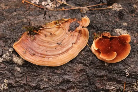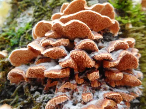
Ciri dan struktur
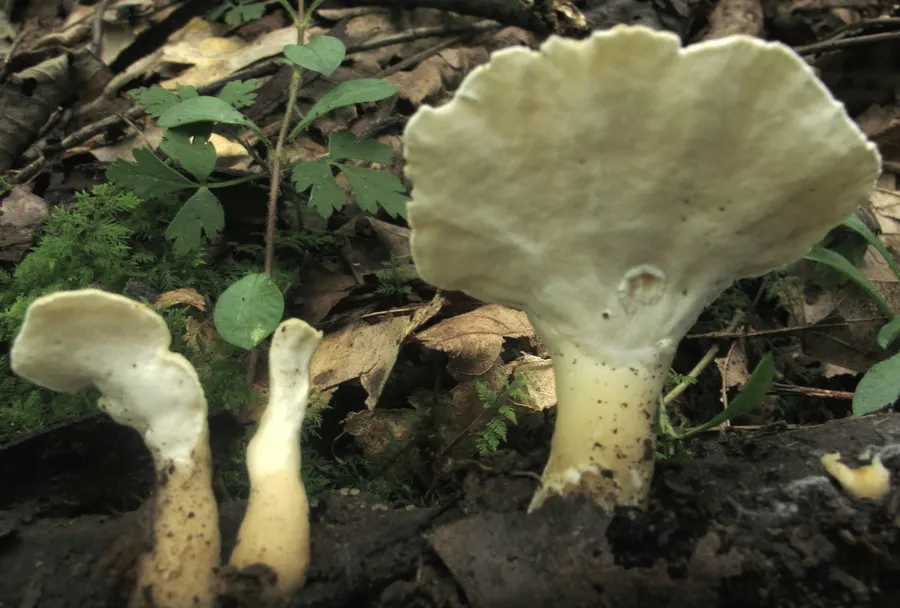
The fruit bodies of Loweomyces fractipes can be quite variable in form. The stipe is placed centrally to laterally, dimidiate with fan- to kidney-shaped caps or almost effused-reflexed, 1–4 cm wide, 1–5 mm thick, soft when fresh, brittle when dry. The upper surface of the cap is white in young specimens, but becomes yellowish with age, at first finely tomentose, with age more adpressed and semi-glabrous, often somewhat wrinkled, usually azonate. When the stipe is present it is white to yellowish, measuring up to 4 cm long, and it is cylindric to flattened and expanded towards the cap. The colour of the pore surface is white to cream, and consists of tiny, angular pores numbering 4–5 per millimetre. The context in cap and stipe are white and comprise two layers: a hard inner or lower layer that is covered with a much looser layer, which may be agglutinated on the surface with age. The tube layer is the same colour as the pore surface, and up to 3 mm thick. The hyphal system is monomitic, containing only generative hyphae. These hyphae have clamp connections, which in the subhymenium and trama are thin-walled and 3–5 μm in diameter. In the context and especially the stipe, the hyphae are much more thick-walled and reminiscent of skeletal hyphae, but with scattered clamps. These hyphae are interpreted by Ryvarden as sclerified generative hyphae. Cystidia are variably present in the hymenium. They are often difficult to observe during microscopy. Their shape is ventricose to cylindrical, and they are thin-walled, measuring 15–25 by 5–6.5 μm. The basidia are broadly club-shaped, and have four sterigmata.
Habitat dan peran ekologis
Loweomyces fractipes is found in Europe, North America, Costa Rica, South America (Brazil), and Korea.
- Habitat atau konteks temuan
- Informasi belum tersedia pada sumber rilis ini.
- Substrat
- Informasi belum tersedia pada sumber rilis ini.
- Peran atau konteks
- Belum dikategorikan pada rilis ini.
Reproduksi
The hyphal system is monomitic, containing only generative hyphae. These hyphae have clamp connections, which in the subhymenium and trama are thin-walled and 3–5 μm in diameter. In the context and especially the stipe, the hyphae are much more thick-walled and reminiscent of skeletal hyphae, but with scattered clamps. These hyphae are interpreted by Ryvarden as sclerified generative hyphae. Cystidia are variably present in the hymenium. They are often difficult to observe during microscopy. Their shape is ventricose to cylindrical, and they are thin-walled, measuring 15–25 by 5–6.5 μm. The basidia are broadly club-shaped, and have four sterigmata. They have a basal clamp, and measure 15–20 by 6–9 μm.
Catatan identitas dan cakupan
- Nama diterima mengikuti Catalogue of Life Base2026-09-11; uraian mempertahankan konsep dan nama sumber aslinya.
- Original geographic, specimen and life-cycle scope retained; source import awaits individual editorial review.
- Impor dan terjemahan otomatis belum diperiksa editorial per profil. Struktur reproduktif yang disebutkan tidak membuktikan seluruh siklus hidup diketahui.
- Pengukuran pada uraian mengikuti spesimen, kultur, dan metode sumber; angka tidak diperluas ke semua populasi.
Catatan distribusi dalam narasi mengikuti sumbernya. Dokumentasi spesimen atau foto mengikuti catatan sumber. Bukti wilayah Indonesia dan penilaian konservasi tidak disimpulkan dari gambar.
Melihat dengan konteks
Dokumentasi Loweomyces fractipes menurut asosiasi foto pada record takson iNaturalist. Kredit: mycowalt. Identitas mengikuti sumber; bukan penentuan spesimen independen.
- Jenis
- Fotografi
- Kredit
- (c) mycowalt, some rights reserved (CC BY-SA), uploaded by mycowalt
- Lisensi
- CC BY-SA 4.0
- Sumber media
- Buka entri sumber
Sumber profil
- [1] Loweomyces fractipes — Wikipedia (en)
- [2] Catalogue of Life Base2026-09-11 — Loweomyces fractipes
- [3] Dokumentasi visual — Loweomyces fractipes
Rujukan halaman ini
Fungi. Loweomyces fractipes. Rilis data fungi-2026-10-12-expansion-2. https://fungi-atlas.mgadib.chatgpt.site/jamur/loweomyces-fractipes/.
Status: impor sumber; pemeriksaan editorial per profil belum selesai. Untuk klaim biologis, gunakan rujukan pada bagian terkait dan periksa konteks sumber aslinya.