01 / Morfologi

Ciri dan struktur

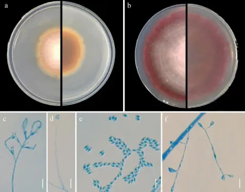
Mariannaea elegans is an anamorphic fungus (i.e., it reproduces asexually). When M. elegans is grown on a Petri dish in 2% MEA (Malt Extract Agar; medium used in a Petri dish) and PDA (potato-dextrose agar) at 22 C the growth and morphological characteristics listed below are observed. The colonies of M. elegans grows better in MEA than in PDA. On average colonies of M. elegans can be observed with the naked eye having a diameter of 2.5–6 cm after 10 days of growth. They appear thin and velvety (i.e. smooth) or floccose (i.e. woolly) or matted (i.e. powdery) and are odourless. Sometimes the colonies make a ring shaped pattern as might be seen on a jawbreaker candy. Specimens isolated from wood possess brown colonies whose brown pigment fuses into the surrounding agar. However, specimens isolated from soil possess reddish-purple colonies whose pigmentation does not diffuse in to the agar surrounding the colonies of M. elegans. Colonies are white to cinnamon buff (peanut butter colour) and possess either smooth or rough walls. The mycelium is either smooth-walled or rough-walled, white to olivaceous (the green colour of the mould on blue cheese) or pink. Conidiophores are arise from mycelia immersed in the agar. They are light brown, with a smooth or verrucose (i.e., bumpy) surface and grow to a maximum of 1000 μm long and reach a maximum of 10 μm in width at the base. Conidiophores can also arise from aerial hyphae. These conidiophores grow to 250 μm in length and 7 μm in width. Additionally, they can grow to reach a maximum of 1400 μm in length and 23 μm in width at their base.

Sebagian uraian ditampilkan dalam bahasa sumber; terjemahannya belum tersedia atau belum lolos pemeriksaan.

Rujukan: [1]

Pengukuran mengikuti objek, spesimen atau kultur, dan metode yang disebutkan sumber. Baca uraian asli untuk konteks lengkap.

02 / Kehidupan

Habitat dan peran ekologis

Mariannaea elegans grows on decaying coniferous bark or wood, forest soil, house dust, prairie and grassland soils, calcareous soil, running water that has little pollution, waste stabilization ponds, acid mine drainage stream, fields treated with digested sewage sludge, in arable soils, and on pine chips. Mariannaea elegans has been isolated from various regions in Canada: Quebec, Ontario, Manitoba, Saskatchewan, and British Columbia. Both varieties have both been recorded in Germany, the Netherlands, the United States, and Canada. However, only M. elegans var. elegans has also be found in France, the British Isles, Italy, Poland, and South Africa, whereas, M. elegans var. punicea has exclusively be found in Democratic Republic of the Congo. The ecological role of this species is not well known. It is involved in wood biodeterioration either through the formation of soft rot cavities or through cell wall erosion. It may also influence the growth of other fungi. For example, at 25 C it inhibits mycelial growth in the mushroom, Pholiota microspora; but at12 C it enhances mycelial growth of P. microspora. It is capable of preventing sapstain (a blue to greyish-black colour) formation on wood treated with M. elegans.

Sebagian uraian ditampilkan dalam bahasa sumber; terjemahannya belum tersedia atau belum lolos pemeriksaan.

Rujukan: [1]

Habitat atau konteks temuan
hutan; padang rumput
Substrat
kayu
Peran atau konteks
Belum dikategorikan pada rilis ini.
Temukan hubungan serupa
03 / Siklus hidup

Reproduksi

Mariannaea elegans is an anamorphic fungus (i.e., it reproduces asexually). When M. elegans is grown on a Petri dish in 2% MEA (Malt Extract Agar; medium used in a Petri dish) and PDA (potato-dextrose agar) at 22 C the growth and morphological characteristics listed below are observed. The colonies of M. elegans grows better in MEA than in PDA. On average colonies of M. elegans can be observed with the naked eye having a diameter of 2.5–6 cm after 10 days of growth. They appear thin and velvety (i.e. smooth) or floccose (i.e. woolly) or matted (i.e. powdery) and are odourless. Sometimes the colonies make a ring shaped pattern as might be seen on a jawbreaker candy. Specimens isolated from wood possess brown colonies whose brown pigment fuses into the surrounding agar.

Sebagian uraian ditampilkan dalam bahasa sumber; terjemahannya belum tersedia atau belum lolos pemeriksaan.

Rujukan: [1]

Sinonim menurut acuan taksonomi rilis: Hormodendrum elegans; Paecilomyces elegans; Penicillium elegans; Spicaria elegans
04 / Batas informasi

Catatan identitas dan cakupan

  • Nama diterima mengikuti Catalogue of Life Base2026-09-11; uraian mempertahankan konsep dan nama sumber aslinya.
  • Original geographic, specimen and life-cycle scope retained; source import awaits individual editorial review.
  • Impor dan terjemahan otomatis belum diperiksa editorial per profil. Struktur reproduktif yang disebutkan tidak membuktikan seluruh siklus hidup diketahui.
  • Pengukuran pada uraian mengikuti spesimen, kultur, dan metode sumber; angka tidak diperluas ke semua populasi.

Catatan distribusi dalam narasi mengikuti sumbernya. Dokumentasi spesimen atau foto mengikuti catatan sumber. Bukti wilayah Indonesia dan penilaian konservasi tidak disimpulkan dari gambar.

05 / Dokumentasi

Melihat dengan konteks

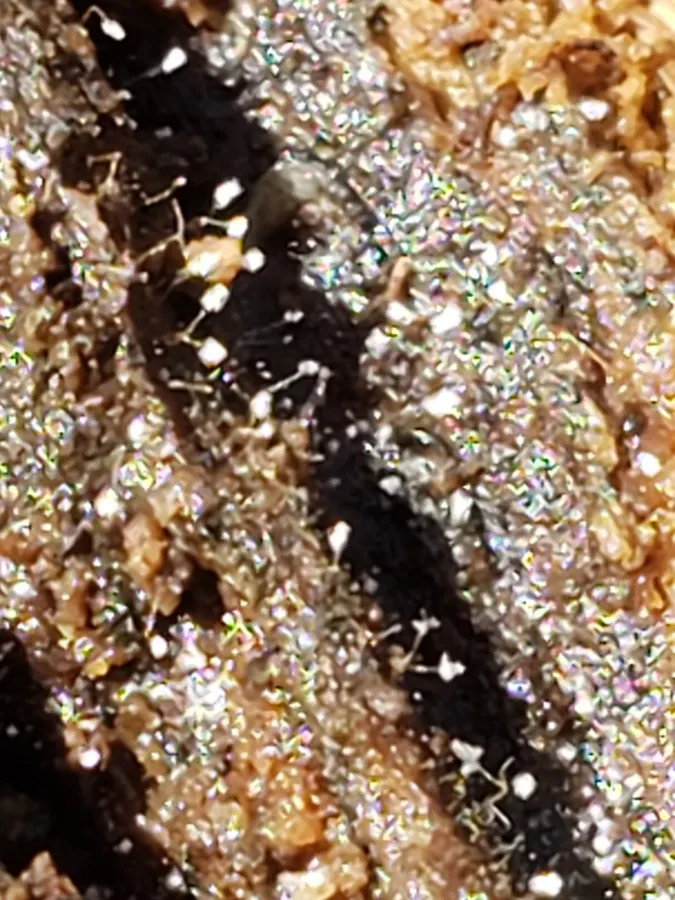
Dokumentasi Mariannaea elegans menurut asosiasi foto pada record takson iNaturalist. Kredit: Mark Richman. Identitas mengikuti sumber; bukan penentuan spesimen independen.

Jenis
Fotografi
Kredit
(c) Mark Richman, some rights reserved (CC BY), uploaded by Mark Richman
Lisensi
CC BY 4.0
Sumber media
Buka entri sumber

Gambar menunjukkan objek menurut identifikasi sumber, bukan verifikasi independen spesimen oleh Fungi. Tidak ada skala baru yang ditambahkan.

06 / Rujukan

Sumber profil

  1. [1] Mariannaea elegans — Wikipedia (en)Bagian sumber: Frozen revision 1334454440
    Tanggal penghimpunan sumber: 2026-10-12
    Teks sumber: CC BY-SA4.0 · Wikipedia contributors
  2. [2] Catalogue of Life Base2026-09-11 — Mariannaea elegansBagian sumber: ColDP NameUsage.tsv ID 3Y674; accepted species; source 2073
    Tanggal penghimpunan sumber: 2026-10-12
  3. [3] Dokumentasi visual — Mariannaea elegansBagian sumber: Individual media record; identification follows the source, not independent determination
    Tanggal penghimpunan sumber: 2026-10-12
    Media sumber: CC BY 4.0 · (c) Mark Richman, some rights reserved (CC BY), uploaded by Mark Richman

Adaptasi teks: CC BY-SA 4.0. Wikipedia contributors — Mariannaea elegans — Wikipedia (en)

Uraian asli dan riwayat sumber

Riwayat atau halaman sumber

Ringkasan sumber

Mariannaea elegans is an anamorphic fungus (i.e., it reproduces asexually). When M. elegans is grown on a Petri dish in 2% MEA (Malt Extract Agar; medium used in a Petri dish) and PDA (potato-dextrose agar) at 22 C the growth and morphological characteristics listed below are observed. The colonies of M. elegans grows better in MEA than in PDA. On average colonies of M. elegans can be observed with the naked eye having a diameter of 2.5–6 cm after 10 days of growth. They appear thin and velvety (i.e. smooth) or floccose (i.e. woolly) or matted (i.e. powdery) and are odourless.

Revisi sumber

Morfologi sumber

Mariannaea elegans is an anamorphic fungus (i.e., it reproduces asexually). When M. elegans is grown on a Petri dish in 2% MEA (Malt Extract Agar; medium used in a Petri dish) and PDA (potato-dextrose agar) at 22 C the growth and morphological characteristics listed below are observed. The colonies of M. elegans grows better in MEA than in PDA. On average colonies of M. elegans can be observed with the naked eye having a diameter of 2.5–6 cm after 10 days of growth. They appear thin and velvety (i.e. smooth) or floccose (i.e. woolly) or matted (i.e. powdery) and are odourless. Sometimes the colonies make a ring shaped pattern as might be seen on a jawbreaker candy. Specimens isolated from wood possess brown colonies whose brown pigment fuses into the surrounding agar. However, specimens isolated from soil possess reddish-purple colonies whose pigmentation does not diffuse in to the agar surrounding the colonies of M. elegans. Colonies are white to cinnamon buff (peanut butter colour) and possess either smooth or rough walls. The mycelium is either smooth-walled or rough-walled, white to olivaceous (the green colour of the mould on blue cheese) or pink. Conidiophores are arise from mycelia immersed in the agar. They are light brown, with a smooth or verrucose (i.e., bumpy) surface and grow to a maximum of 1000 μm long and reach a maximum of 10 μm in width at the base. Conidiophores can also arise from aerial hyphae. These conidiophores grow to 250 μm in length and 7 μm in width. Additionally, they can grow to reach a maximum of 1400 μm in length and 23 μm in width at their base.

Revisi sumber

Ekologi sumber

Mariannaea elegans grows on decaying coniferous bark or wood, forest soil, house dust, prairie and grassland soils, calcareous soil, running water that has little pollution, waste stabilization ponds, acid mine drainage stream, fields treated with digested sewage sludge, in arable soils, and on pine chips. Mariannaea elegans has been isolated from various regions in Canada: Quebec, Ontario, Manitoba, Saskatchewan, and British Columbia. Both varieties have both been recorded in Germany, the Netherlands, the United States, and Canada. However, only M. elegans var. elegans has also be found in France, the British Isles, Italy, Poland, and South Africa, whereas, M. elegans var. punicea has exclusively be found in Democratic Republic of the Congo. The ecological role of this species is not well known. It is involved in wood biodeterioration either through the formation of soft rot cavities or through cell wall erosion. It may also influence the growth of other fungi. For example, at 25 C it inhibits mycelial growth in the mushroom, Pholiota microspora; but at12 C it enhances mycelial growth of P. microspora. It is capable of preventing sapstain (a blue to greyish-black colour) formation on wood treated with M. elegans.

Revisi sumber

Struktur reproduksi sumber

Mariannaea elegans is an anamorphic fungus (i.e., it reproduces asexually). When M. elegans is grown on a Petri dish in 2% MEA (Malt Extract Agar; medium used in a Petri dish) and PDA (potato-dextrose agar) at 22 C the growth and morphological characteristics listed below are observed. The colonies of M. elegans grows better in MEA than in PDA. On average colonies of M. elegans can be observed with the naked eye having a diameter of 2.5–6 cm after 10 days of growth. They appear thin and velvety (i.e. smooth) or floccose (i.e. woolly) or matted (i.e. powdery) and are odourless. Sometimes the colonies make a ring shaped pattern as might be seen on a jawbreaker candy. Specimens isolated from wood possess brown colonies whose brown pigment fuses into the surrounding agar.

Revisi sumber

Rujukan halaman ini

Fungi. Mariannaea elegans. Rilis data fungi-2026-10-12-expansion-2. https://fungi-atlas.mgadib.chatgpt.site/jamur/mariannaea-elegans/.

Status: impor sumber; pemeriksaan editorial per profil belum selesai. Untuk klaim biologis, gunakan rujukan pada bagian terkait dan periksa konteks sumber aslinya.