01 / Morfologi

Ciri dan struktur

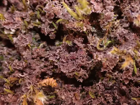
M. patagonica (Fig. 3) is morphologically similar to M. carnosa. Generally, spore characters are the best distinguishing characters (Fig. 4). The spores of M. carnosa are longer and 92% of 72 measured spores were in the range 23–35 µm. By contrast, 70% of 58 measured spores of M. patagonica were in the range 15–22 µm. This means that there is an overlap in sizes between these two species. However, the spores of M. patagonica are often two-septate, sometimes three-septate, which is very rare in M. carnosa. Here follows a more detailed treatment of Massalongia patagonica Kitaura & Lorenz: Thallus foliose, forming rosettes up to 3 cm, mostly muscicolous; lobes 0.5–1.5 mm broad, up to 1 cm long, irregularly and repeatedly divided with isidioid marginal outgrowths, simple to sparingly branched, sometimes developing into branched lobule systems, lobules, 0.1 mm wide. Upper surface brown, glabrous and glossy; upper cortex 20–30 µm thick, paraplectenchymatic, of thick-walled (ca. 1.5–2 µm wide) cells with 7–12 µm large lumina; photobiont layer 40–60 µm thick, often also developed in the subhymenium; cyanobiont Nostoc, cells bluish-green, irregularly subglobose to ellipsoid, 5–9 × 6–11 µm in size, arranged within 20–40 µm large glomeruli without visible chain structures, chain structures visible in some liberated cells; medulla loose, 60–80 µm thick; lower cortex absent, with scattered rhizohyphae. Apothecia common to scattered, substipitate, laminal, 1–2 mm wide; thalline excipulum lacking, true excipulum weakly prominent; epithecium 5–10 µm thick, of protruding brown and strongly swollen, pyriform paraphyse end cells, 4–6 µm wide, 7–10 µm long, paraphyses undivided to sparingly divided, 2–4 µm thick; hymenium ca. 60 µm thick, IKI + blue; asci clavate 50–70 × 10–15 µm, 8-spored, with distinct internal apical IKI + blue sheath-like structures, sometimes also with weak tube structures; ascospores narrowly ellipsoid, occasionally asymmetric, 1- to 2 (3)-septate, (13) 20–25 (28) × 5–7.5 µm.

Sebagian uraian ditampilkan dalam bahasa sumber; terjemahannya belum tersedia atau belum lolos pemeriksaan.

Rujukan: [1]

Pengukuran mengikuti objek, spesimen atau kultur, dan metode yang disebutkan sumber. Baca uraian asli untuk konteks lengkap.

02 / Kehidupan

Habitat dan peran ekologis

This is a species of wet to dry rock surfaces or boulders, usually growing in between mosses or on plant remains. It has a widely scattered distribution in South America, ranging from the temperate forests of south-central Chile, including the Juan Fernandez Islands and Patagonia, with two widely separated collections from southernmost Chile and Argentinean Tierra del Fuego. In addition, it is known from the Falkland Islands, Antarctica, mountains of SE Australia, where it is rare and from several localities in New Zealand.

Sebagian uraian ditampilkan dalam bahasa sumber; terjemahannya belum tersedia atau belum lolos pemeriksaan.

Rujukan: [1]

Habitat atau konteks temuan
hutan
Substrat
Informasi belum tersedia pada sumber rilis ini.
Peran atau konteks
Pembentuk liken
Temukan hubungan serupa
03 / Siklus hidup

Reproduksi

M. patagonica (Fig. 3) is morphologically similar to M. carnosa. Generally, spore characters are the best distinguishing characters (Fig. 4). The spores of M. carnosa are longer and 92% of 72 measured spores were in the range 23–35 µm. By contrast, 70% of 58 measured spores of M. patagonica were in the range 15–22 µm. This means that there is an overlap in sizes between these two species. However, the spores of M. patagonica are often two-septate, sometimes three-septate, which is very rare in M. carnosa. Here follows a more detailed treatment of Massalongia patagonica Kitaura & Lorenz: Thallus foliose, forming rosettes up to 3 cm, mostly muscicolous; lobes 0.5–1.5 mm broad, up to 1 cm long, irregularly and repeatedly divided with isidioid marginal outgrowths, simple to sparingly branched, sometimes developing into branched lobule systems, lobules, 0.1 mm wide.

Sebagian uraian ditampilkan dalam bahasa sumber; terjemahannya belum tersedia atau belum lolos pemeriksaan.

Rujukan: [1]

04 / Batas informasi

Catatan identitas dan cakupan

  • Nama diterima mengikuti Catalogue of Life Base2026-09-11; uraian mempertahankan konsep dan nama sumber aslinya.
  • Preserve original treatment localities, hosts and substrate scope. Materials examined describe documented specimens, not the full global range.
  • Impor dan terjemahan otomatis belum diperiksa editorial per profil. Struktur reproduktif yang disebutkan tidak membuktikan seluruh siklus hidup diketahui.
  • Pengukuran pada uraian mengikuti spesimen, kultur, dan metode sumber; angka tidak diperluas ke semua populasi.

Catatan distribusi dalam narasi mengikuti sumbernya. Dokumentasi spesimen atau foto mengikuti catatan sumber. Bukti wilayah Indonesia dan penilaian konservasi tidak disimpulkan dari gambar.

05 / Dokumentasi

Melihat dengan konteks

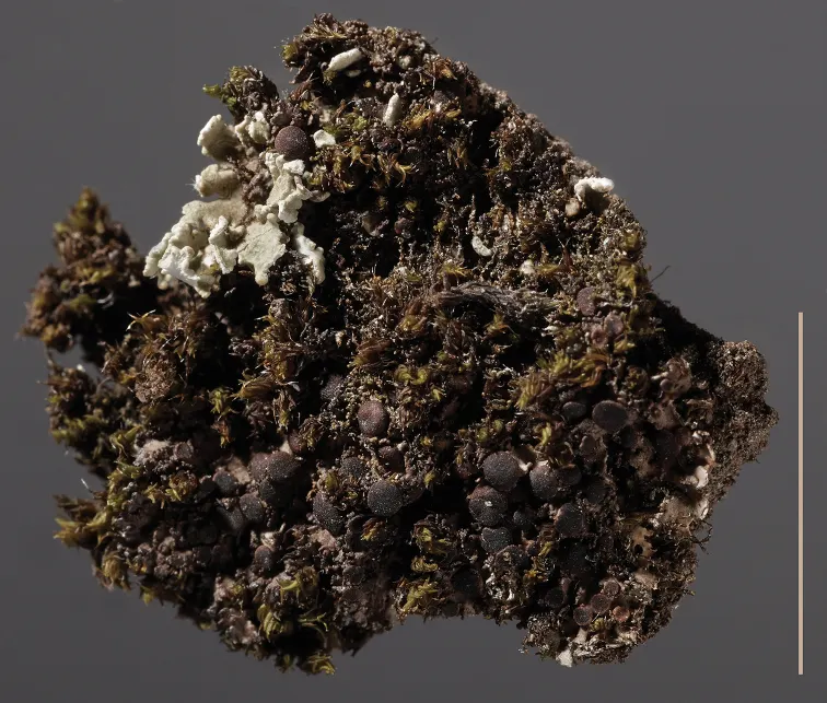
Massalongia patagonica, AE 15-033. Scale bar: 1 cm.

Jenis
Pelat ilmiah
Kredit
Per M. Jørgensen, Heidi L. Andersen, Arve Elvebakk
Lisensi
CC BY 4.0
Sumber media
Buka entri sumber

Gambar menunjukkan objek menurut identifikasi sumber, bukan verifikasi independen spesimen oleh Fungi. Tidak ada skala baru yang ditambahkan.

06 / Rujukan

Sumber profil

  1. [1] The genus Massalongia (lichenised ascomycetae) in the Southern Hemisphere — Massalongia patagonicaBagian sumber: PMC6906167_section_8
    Tanggal penghimpunan sumber: 2026-10-12
    Teks sumber: CC BY 4.0 · Jørgensen PM, Andersen HL, Elvebakk A.
  2. [2] Catalogue of Life Base2026-09-11 — Massalongia patagonicaBagian sumber: ColDP NameUsage.tsv ID 3YBTG; accepted species; source 2073
    Tanggal penghimpunan sumber: 2026-10-12
  3. [3] Dokumentasi visual — Massalongia patagonicaBagian sumber: Individual media record; identification follows the source, not independent determination
    Tanggal penghimpunan sumber: 2026-10-12
    Media sumber: CC BY 4.0 · Per M. Jørgensen, Heidi L. Andersen, Arve Elvebakk

Adaptasi teks: CC BY 4.0. Jørgensen PM, Andersen HL, Elvebakk A. — The genus Massalongia (lichenised ascomycetae) in the Southern Hemisphere — Massalongia patagonica

Uraian asli dan riwayat sumber

Riwayat atau halaman sumber

Ringkasan sumber

M. patagonica (Fig. 3) is morphologically similar to M. carnosa. Generally, spore characters are the best distinguishing characters (Fig. 4). The spores of M. carnosa are longer and 92% of 72 measured spores were in the range 23–35 µm. By contrast, 70% of 58 measured spores of M. patagonica were in the range 15–22 µm. This means that there is an overlap in sizes between these two species. However, the spores of M. patagonica are often two-septate, sometimes three-septate, which is very rare in M. carnosa.

Revisi sumber

Morfologi sumber

M. patagonica (Fig. 3) is morphologically similar to M. carnosa. Generally, spore characters are the best distinguishing characters (Fig. 4). The spores of M. carnosa are longer and 92% of 72 measured spores were in the range 23–35 µm. By contrast, 70% of 58 measured spores of M. patagonica were in the range 15–22 µm. This means that there is an overlap in sizes between these two species. However, the spores of M. patagonica are often two-septate, sometimes three-septate, which is very rare in M. carnosa. Here follows a more detailed treatment of Massalongia patagonica Kitaura & Lorenz: Thallus foliose, forming rosettes up to 3 cm, mostly muscicolous; lobes 0.5–1.5 mm broad, up to 1 cm long, irregularly and repeatedly divided with isidioid marginal outgrowths, simple to sparingly branched, sometimes developing into branched lobule systems, lobules, 0.1 mm wide. Upper surface brown, glabrous and glossy; upper cortex 20–30 µm thick, paraplectenchymatic, of thick-walled (ca. 1.5–2 µm wide) cells with 7–12 µm large lumina; photobiont layer 40–60 µm thick, often also developed in the subhymenium; cyanobiont Nostoc, cells bluish-green, irregularly subglobose to ellipsoid, 5–9 × 6–11 µm in size, arranged within 20–40 µm large glomeruli without visible chain structures, chain structures visible in some liberated cells; medulla loose, 60–80 µm thick; lower cortex absent, with scattered rhizohyphae. Apothecia common to scattered, substipitate, laminal, 1–2 mm wide; thalline excipulum lacking, true excipulum weakly prominent; epithecium 5–10 µm thick, of protruding brown and strongly swollen, pyriform paraphyse end cells, 4–6 µm wide, 7–10 µm long, paraphyses undivided to sparingly divided, 2–4 µm thick; hymenium ca. 60 µm thick, IKI + blue; asci clavate 50–70 × 10–15 µm, 8-spored, with distinct internal apical IKI + blue sheath-like structures, sometimes also with weak tube structures; ascospores narrowly ellipsoid, occasionally asymmetric, 1- to 2 (3)-septate, (13) 20–25 (28) × 5–7.5 µm.

Revisi sumber

Ekologi sumber

This is a species of wet to dry rock surfaces or boulders, usually growing in between mosses or on plant remains. It has a widely scattered distribution in South America, ranging from the temperate forests of south-central Chile, including the Juan Fernandez Islands and Patagonia, with two widely separated collections from southernmost Chile and Argentinean Tierra del Fuego. In addition, it is known from the Falkland Islands, Antarctica, mountains of SE Australia, where it is rare and from several localities in New Zealand.

Revisi sumber

Struktur reproduksi sumber

M. patagonica (Fig. 3) is morphologically similar to M. carnosa. Generally, spore characters are the best distinguishing characters (Fig. 4). The spores of M. carnosa are longer and 92% of 72 measured spores were in the range 23–35 µm. By contrast, 70% of 58 measured spores of M. patagonica were in the range 15–22 µm. This means that there is an overlap in sizes between these two species. However, the spores of M. patagonica are often two-septate, sometimes three-septate, which is very rare in M. carnosa. Here follows a more detailed treatment of Massalongia patagonica Kitaura & Lorenz: Thallus foliose, forming rosettes up to 3 cm, mostly muscicolous; lobes 0.5–1.5 mm broad, up to 1 cm long, irregularly and repeatedly divided with isidioid marginal outgrowths, simple to sparingly branched, sometimes developing into branched lobule systems, lobules, 0.1 mm wide.

Revisi sumber

Rujukan halaman ini

Fungi. Massalongia patagonica. Rilis data fungi-2026-10-12-expansion-2. https://fungi-atlas.mgadib.chatgpt.site/jamur/massalongia-patagonica/.

Status: impor sumber; pemeriksaan editorial per profil belum selesai. Untuk klaim biologis, gunakan rujukan pada bagian terkait dan periksa konteks sumber aslinya.