Ciri dan struktur
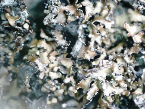
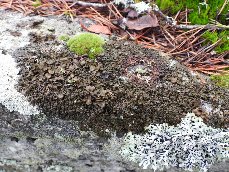
M. stygia has foliose thallus, composed of 0.25–1.5 mm broad, smooth and usually distinctly convex lobes (Szczepańska and Kossowska 2017). The upper surface is glossy, dark brown to almost black. The lower surface is dark brown to black, paler near the margins, with single, dark rhizines. Pseudocyphellae in this species are numerous, rounded or slightly elongated and laminal – clearly visible on the upper surface of the lobes. Pycnidia are also common, globose, laminal and immersed with hyaline, bacilliform conidia (3.5–5 × 1 µm). Apothecia are laminal, constricted at the base and 0.5–6 mm in diameter. Ascospores are hyaline, ellipsoid to oblong-ellipsoid, 6–8 × 4–6 μm in size.
Habitat dan peran ekologis
M. stygia is a circumpolar and arctic-alpine species occurring mainly on siliceous rocks in upper mountain areas in North America and Europe (Otte et al. 2005). Available molecular data concern only a few samples collected in North America (Greenland) and North (Iceland, Finland, Norway) and West (Italy) Europe.
- Habitat atau konteks temuan
- Informasi belum tersedia pada sumber rilis ini.
- Substrat
- Informasi belum tersedia pada sumber rilis ini.
- Peran atau konteks
- Pembentuk liken
Reproduksi
M. stygia has foliose thallus, composed of 0.25–1.5 mm broad, smooth and usually distinctly convex lobes (Szczepańska and Kossowska 2017). The upper surface is glossy, dark brown to almost black. The lower surface is dark brown to black, paler near the margins, with single, dark rhizines. Pseudocyphellae in this species are numerous, rounded or slightly elongated and laminal – clearly visible on the upper surface of the lobes. Pycnidia are also common, globose, laminal and immersed with hyaline, bacilliform conidia (3.5–5 × 1 µm). Apothecia are laminal, constricted at the base and 0.5–6 mm in diameter. Ascospores are hyaline, ellipsoid to oblong-ellipsoid, 6–8 × 4–6 μm in size.
Catatan identitas dan cakupan
- Nama diterima mengikuti Catalogue of Life Base2026-09-11; uraian mempertahankan konsep dan nama sumber aslinya.
- Preserve original treatment localities, hosts and substrate scope. Materials examined describe documented specimens, not the full global range.
- Impor dan terjemahan otomatis belum diperiksa editorial per profil. Struktur reproduktif yang disebutkan tidak membuktikan seluruh siklus hidup diketahui.
- Pengukuran pada uraian mengikuti spesimen, kultur, dan metode sumber; angka tidak diperluas ke semua populasi.
Catatan distribusi dalam narasi mengikuti sumbernya. Dokumentasi spesimen atau foto mengikuti catatan sumber. Bukti wilayah Indonesia dan penilaian konservasi tidak disimpulkan dari gambar.
Melihat dengan konteks
Dokumentasi Melanelia stygia menurut asosiasi foto pada record takson iNaturalist. Kredit: Vesa Oikonen. Identitas mengikuti sumber; bukan penentuan spesimen independen.
- Jenis
- Fotografi
- Kredit
- no rights reserved, uploaded by Vesa Oikonen
- Lisensi
- CC0 1.0
- Sumber media
- Buka entri sumber
Dokumentasi tambahan
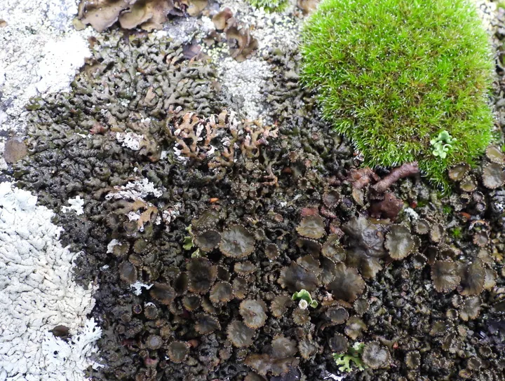
Foto yang diasosiasikan dengan Melanelia stygia pada iNaturalist. Identifikasi mengikuti sumber; bukan penentuan spesimen independen.
Kredit dan penggunaan
no rights reserved, uploaded by Vesa Oikonen
Ukuran dan format gambar disesuaikan tanpa pemotongan, retus, atau penambahan skala. Lisensi sumber dipertahankan.
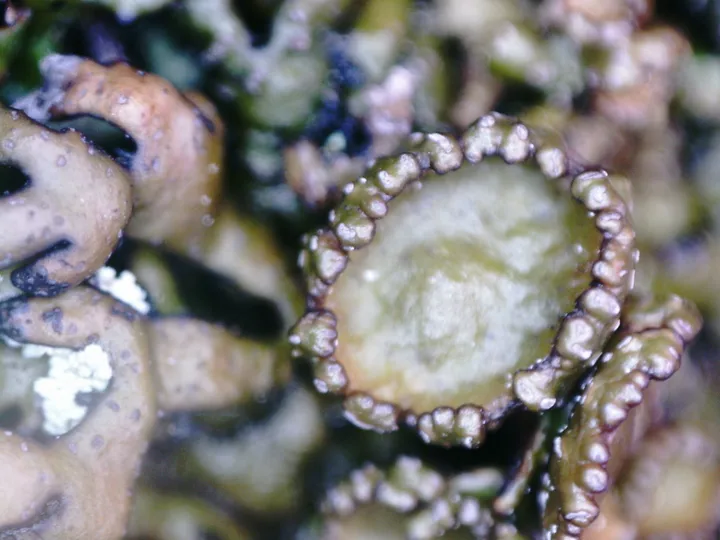
Foto yang diasosiasikan dengan Melanelia stygia pada iNaturalist. Identifikasi mengikuti sumber; bukan penentuan spesimen independen.
Kredit dan penggunaan
no rights reserved, uploaded by Vesa Oikonen
Ukuran dan format gambar disesuaikan tanpa pemotongan, retus, atau penambahan skala. Lisensi sumber dipertahankan.
Sumber profil
- [1] Infraspecific variation of some brown Parmeliae (in Poland) – a comparison of ITS rDNA and non-molecular characters — Melanelia stygia
- [2] Catalogue of Life Base2026-09-11 — Melanelia stygia
- [3] Dokumentasi visual — Melanelia stygia
- [4] Dokumentasi visual — Melanelia stygia
- [5] Dokumentasi visual — Melanelia stygia
Rujukan halaman ini
Fungi. Melanelia stygia. Rilis data fungi-2026-10-12-expansion-2. https://fungi-atlas.mgadib.chatgpt.site/jamur/melanelia-stygia/.
Status: impor sumber; pemeriksaan editorial per profil belum selesai. Untuk klaim biologis, gunakan rujukan pada bagian terkait dan periksa konteks sumber aslinya.