Ciri dan struktur
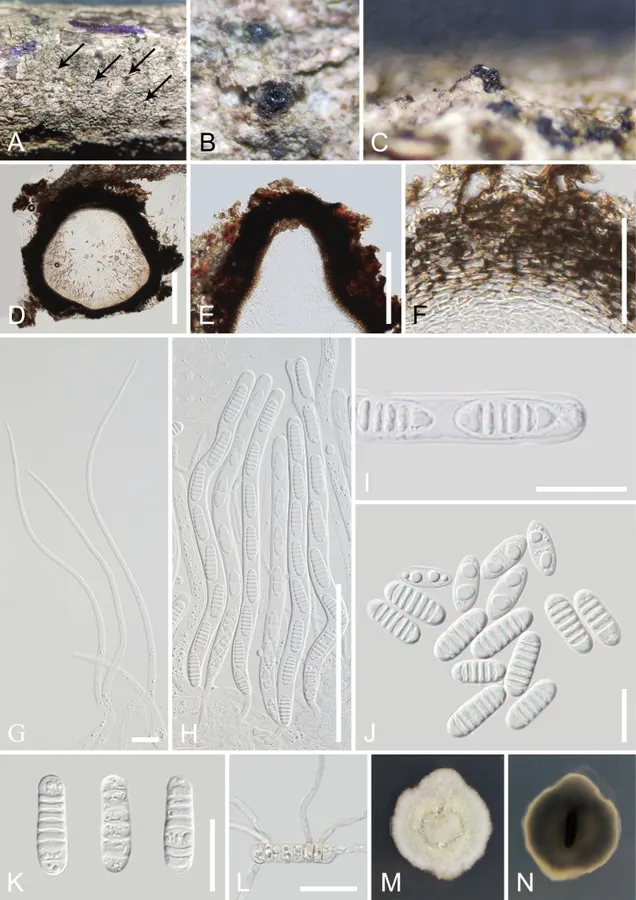
Saprobic on a dead branch of Aquilaria sp. Sexual morph: Ascomata (excluding neck) 400–600 µm high × 430–580 µm diam. (x– = 515 × 520 µm, n = 10), solitary, scattered to gregarious, semi-immersed to immersed, erumpent through host tissue, globose to subglobose, black, coriaceous to carbonaceous, ostiolate. Ostiolar canal 230–365 µm high × 200–260 µm wide (x– = 303 × 230 µm, n = 10), central, black, conical, coriaceous to carbonaceous, filled with hyaline sparse periphyses. Peridium 30–120 µm wide (x– = 75 µm, n = 20), comprising dense, several layers of thick-walled cells of textura angularis to textura prismatica, outer layers brown to dark brown, becoming lighter inwardly. Hamathecium comprising 2.5–6.5 µm wide, numerous filamentous, filiform, septate, unbranched, hyaline pseudoparaphyses, attached to the base and between the asci, embedded in a gelatinous matrix. Asci 175–220 × 8.5–11.5 µm (x– = 195 × 10.5 µm, n = 30), bitunicate, 8-spored, cylindrical, long pedicel, thickened and rounded apex, with an obvious ocular chamber. Ascospores (17.5–)20–26.5 × 7–9 µm (x– = 24 × 8 µm, n = 30), overlapping-uniseriate, hyaline, when ascospores gather together, they appear light yellow, mostly 6–7-septate at maturity, cylindrical, with rounded ends, slightly constricted at the septum, often similar width of cells with several small guttules, not surrounded by a mucilaginous sheath. Asexual morph: Undetermined. Ascospores germinated on PDA after 24 hours, germ tubes were produced from most cells, germinated ascospores appear light yellow. Colonies on PDA reaching 3 cm diam., after two weeks at 23–28 °C.
Habitat dan peran ekologis
Aquilaria sp. (China; this study), Camellia sinensis (Thailand; Hyde et al. 2018), and Hevea brasiliensis (Thailand; Senwanna et al. 2021).
- Habitat atau konteks temuan
- Informasi belum tersedia pada sumber rilis ini.
- Substrat
- Informasi belum tersedia pada sumber rilis ini.
- Peran atau konteks
- Belum dikategorikan pada rilis ini.
Reproduksi
Saprobic on a dead branch of Aquilaria sp. Sexual morph: Ascomata (excluding neck) 400–600 µm high × 430–580 µm diam. (x– = 515 × 520 µm, n = 10), solitary, scattered to gregarious, semi-immersed to immersed, erumpent through host tissue, globose to subglobose, black, coriaceous to carbonaceous, ostiolate. Ostiolar canal 230–365 µm high × 200–260 µm wide (x– = 303 × 230 µm, n = 10), central, black, conical, coriaceous to carbonaceous, filled with hyaline sparse periphyses. Peridium 30–120 µm wide (x– = 75 µm, n = 20), comprising dense, several layers of thick-walled cells of textura angularis to textura prismatica, outer layers brown to dark brown, becoming lighter inwardly. Hamathecium comprising 2.5–6.5 µm wide, numerous filamentous, filiform, septate, unbranched, hyaline pseudoparaphyses, attached to the base and between the asci, embedded in a gelatinous matrix.
Catatan identitas dan cakupan
- Nama diterima mengikuti Catalogue of Life Base2026-09-11; uraian mempertahankan konsep dan nama sumber aslinya.
- Preserve original treatment localities, hosts and substrate scope. Materials examined describe documented specimens, not the full global range.
- Impor dan terjemahan otomatis belum diperiksa editorial per profil. Struktur reproduktif yang disebutkan tidak membuktikan seluruh siklus hidup diketahui.
- Pengukuran pada uraian mengikuti spesimen, kultur, dan metode sumber; angka tidak diperluas ke semua populasi.
Catatan distribusi dalam narasi mengikuti sumbernya. Dokumentasi spesimen atau foto mengikuti catatan sumber. Bukti wilayah Indonesia dan penilaian konservasi tidak disimpulkan dari gambar.
Melihat dengan konteks
Melomastia sinensis (GMB-W 1006, new host and geographical record) A–C appearance of ascomata on the host (A the arrows indicate ascomata) D vertical sections through the ascoma E ostiole F peridium G pseudoparaphyses H asci I ascus with an ocular chamber J, K ascospores L germinated ascospore M, N colony on PDA obverse and reverse view. Scale bars: 200 µm (D, E); 100 µm (H); 50 µm (F); 20 µm (I–L); 10 µm (G).
- Jenis
- Pelat ilmiah
- Kredit
- Tian-Ye Du, Samantha C. Karunarathna, Saowaluck Tibpromma, Kevin D. Hyde, Somrudee Nilthong, Ausana Mapook, Xiang-Fu Liu, Dong-Qin Dai, Chen Niu, Abdallah M. Elgorban, Ekachai Chukeatirote, Hao-Han Wang
- Lisensi
- CC BY 4.0
- Sumber media
- Buka entri sumber
Sumber profil
- [1] Melomastia (Dothideomycetes, Ascomycota) species associated with Chinese Aquilaria spp. — Melomastia sinensis
- [2] Catalogue of Life Base2026-09-11 — Melomastia sinensis
- [3] Dokumentasi visual — Melomastia sinensis
Rujukan halaman ini
Fungi. Melomastia sinensis. Rilis data fungi-2026-10-12-expansion-2. https://fungi-atlas.mgadib.chatgpt.site/jamur/melomastia-sinensis/.
Status: impor sumber; pemeriksaan editorial per profil belum selesai. Untuk klaim biologis, gunakan rujukan pada bagian terkait dan periksa konteks sumber aslinya.
