01 / Morfologi

Ciri dan struktur

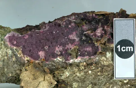
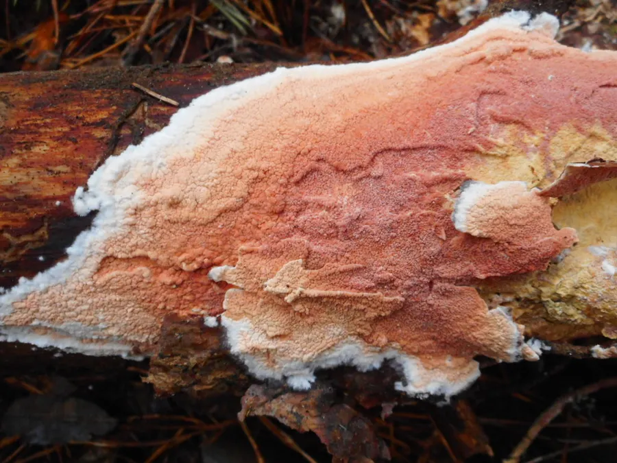
Hymenophore resupinate, annual, loosely attached, effused forming linear areas to 10 x 3 cm; hymenial surface reddish-brown with a purple tinge, becoming vinaceous when old, at first reticulateplicate, becoming porose-reticulate, pores 2-3 per mm, often with a linear arrangement; margin irregular, 1-2 mm wide, byssoid, white, becoming darker with age, loosely attached. Context white, to 400 µm thick, of loosely intertwined hyphae embedding crystals, hyphae in the subhymenium encrusted with orange mucilage granules; generative hyphae 3-4 µm diameter, walls 0.5 µm thick, to 1 µm towards the base, hyaline, without clamp connections. Hymenial layer to 50 µm deep, a dense palisade of basidia, paraphyses, and paraphysate hyphae. Basidia subclavate, 10-14 x 3-4 µm bearing 4 spores; sterigmata slender, erect, to 4 µm long. Paraphyses subclavate, 8-12 x 3-3.5 µm. Paraphysate hyphae filiform, projecting to 25 µm, tinted, 3-4 µm diameter. Spores allantoid, 4-5 x 0.75-1 µm, walls smooth, hyaline, 0.1 µm thick.

Sebagian uraian ditampilkan dalam bahasa sumber; terjemahannya belum tersedia atau belum lolos pemeriksaan.

Rujukan: [1]

Pengukuran mengikuti objek, spesimen atau kultur, dan metode yang disebutkan sumber. Baca uraian asli untuk konteks lengkap.

02 / Kehidupan

Habitat dan peran ekologis

DISTRIBUTION: North America, Australia, Tasmania, New Zealand. HABITAT: Effused on decorticated fallen branches.

Sebagian uraian ditampilkan dalam bahasa sumber; terjemahannya belum tersedia atau belum lolos pemeriksaan.

Rujukan: [1]

Habitat atau konteks temuan
Informasi belum tersedia pada sumber rilis ini.
Substrat
Informasi belum tersedia pada sumber rilis ini.
Peran atau konteks
Belum dikategorikan pada rilis ini.
Temukan hubungan serupa
03 / Siklus hidup

Reproduksi

Hymenophore resupinate, annual, loosely attached, effused forming linear areas to 10 x 3 cm; hymenial surface reddish-brown with a purple tinge, becoming vinaceous when old, at first reticulateplicate, becoming porose-reticulate, pores 2-3 per mm, often with a linear arrangement; margin irregular, 1-2 mm wide, byssoid, white, becoming darker with age, loosely attached. Context white, to 400 µm thick, of loosely intertwined hyphae embedding crystals, hyphae in the subhymenium encrusted with orange mucilage granules; generative hyphae 3-4 µm diameter, walls 0.5 µm thick, to 1 µm towards the base, hyaline, without clamp connections. Hymenial layer to 50 µm deep, a dense palisade of basidia, paraphyses, and paraphysate hyphae.

Sebagian uraian ditampilkan dalam bahasa sumber; terjemahannya belum tersedia atau belum lolos pemeriksaan.

Rujukan: [1]

Sinonim menurut acuan taksonomi rilis: Caloporus taxicola; Gloeoporus taxicola; Gloeoporus taxicola f. longisporus; Merulioporia taxicola; Merulius pulcher; Merulius pulchrum; Merulius ravenelii; Merulius taxicola; Polyporus albocarneus; Polyporus brunneus subsp. rufus; Polyporus brunneus var. rufus; Polyporus haematodes; Polyporus rufus; Polyporus rufus f. lilacina; Polyporus sorbicola; Poria rufa; Poria sorbicola; Poria taxicola; Sesia ravenelii; Xylomyzon pulchrum; Xylomyzon taxicola
04 / Batas informasi

Catatan identitas dan cakupan

  • Nama diterima mengikuti Catalogue of Life Base2026-09-11; uraian mempertahankan konsep dan nama sumber aslinya.
  • Historical description as cited by BiotaNZ. Preserve explicit New Zealand/locality, host and specimen qualifiers. Distribution is the cited source scope, not a current global range.
  • Impor dan terjemahan otomatis belum diperiksa editorial per profil. Struktur reproduktif yang disebutkan tidak membuktikan seluruh siklus hidup diketahui.
  • Pengukuran pada uraian mengikuti spesimen, kultur, dan metode sumber; angka tidak diperluas ke semua populasi.

Catatan distribusi dalam narasi mengikuti sumbernya. Dokumentasi spesimen atau foto mengikuti catatan sumber. Bukti wilayah Indonesia dan penilaian konservasi tidak disimpulkan dari gambar.

05 / Dokumentasi

Melihat dengan konteks

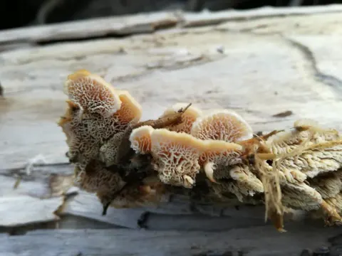
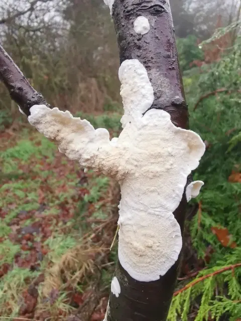
Dokumentasi Meruliopsis taxicola menurut asosiasi foto pada record takson iNaturalist. Kredit: Eugene Popov. Identitas mengikuti sumber; bukan penentuan spesimen independen.

Jenis
Fotografi
Kredit
(c) Eugene Popov, bazı hakları saklıdır (CC BY), yükleyen: Eugene Popov
Lisensi
CC BY 4.0
Sumber media
Buka entri sumber

Gambar menunjukkan objek menurut identifikasi sumber, bukan verifikasi independen spesimen oleh Fungi. Tidak ada skala baru yang ditambahkan.

06 / Rujukan

Sumber profil

  1. [1] Cunningham, G.H. 1963: The Thelephoraceae of Australia and New Zealand. New Zealand Department of Scientific and Industrial Research, Bulletin. 145. — Merulius raveneliiBagian sumber: biota:1cb0defb-36b9-11d5-9548-00d0592d548c:1cb19357-36b9-11d5-9548-00d0592d548c
    Tanggal penghimpunan sumber: 2026-10-12
    Teks sumber: CC BY 4.0 · Cunningham, G.H. 1963: The Thelephoraceae of Australia and New Zealand. New Zealand Department of Scientific and Industrial Research, Bulletin. 145.
  2. [2] Catalogue of Life Base2026-09-11 — Meruliopsis taxicolaBagian sumber: ColDP NameUsage.tsv ID QNMSR; accepted species; source 2073
    Tanggal penghimpunan sumber: 2026-10-12
  3. [3] Dokumentasi visual — Meruliopsis taxicolaBagian sumber: Individual media record; identification follows the source, not independent determination
    Tanggal penghimpunan sumber: 2026-10-12
    Media sumber: CC BY 4.0 · (c) Eugene Popov, bazı hakları saklıdır (CC BY), yükleyen: Eugene Popov

Adaptasi teks: CC BY 4.0. Cunningham, G.H. 1963: The Thelephoraceae of Australia and New Zealand. New Zealand Department of Scientific and Industrial Research, Bulletin. 145. — Cunningham, G.H. 1963: The Thelephoraceae of Australia and New Zealand. New Zealand Department of Scientific and Industrial Research, Bulletin. 145. — Merulius ravenelii

Uraian asli dan riwayat sumber

Riwayat atau halaman sumber

Ringkasan sumber

Hymenophore resupinate, annual, loosely attached, effused forming linear areas to 10 x 3 cm; hymenial surface reddish-brown with a purple tinge, becoming vinaceous when old, at first reticulateplicate, becoming porose-reticulate, pores 2-3 per mm, often with a linear arrangement; margin irregular, 1-2 mm wide, byssoid, white, becoming darker with age, loosely attached. Context white, to 400 µm thick, of loosely intertwined hyphae embedding crystals, hyphae in the subhymenium encrusted with orange mucilage granules; generative hyphae 3-4 µm diameter, walls 0.5 µm thick, to 1 µm towards the base, hyaline, without clamp connections.

Revisi sumber

Morfologi sumber

Hymenophore resupinate, annual, loosely attached, effused forming linear areas to 10 x 3 cm; hymenial surface reddish-brown with a purple tinge, becoming vinaceous when old, at first reticulateplicate, becoming porose-reticulate, pores 2-3 per mm, often with a linear arrangement; margin irregular, 1-2 mm wide, byssoid, white, becoming darker with age, loosely attached. Context white, to 400 µm thick, of loosely intertwined hyphae embedding crystals, hyphae in the subhymenium encrusted with orange mucilage granules; generative hyphae 3-4 µm diameter, walls 0.5 µm thick, to 1 µm towards the base, hyaline, without clamp connections. Hymenial layer to 50 µm deep, a dense palisade of basidia, paraphyses, and paraphysate hyphae. Basidia subclavate, 10-14 x 3-4 µm bearing 4 spores; sterigmata slender, erect, to 4 µm long. Paraphyses subclavate, 8-12 x 3-3.5 µm. Paraphysate hyphae filiform, projecting to 25 µm, tinted, 3-4 µm diameter. Spores allantoid, 4-5 x 0.75-1 µm, walls smooth, hyaline, 0.1 µm thick.

Revisi sumber

Ekologi sumber

DISTRIBUTION: North America, Australia, Tasmania, New Zealand. HABITAT: Effused on decorticated fallen branches.

Revisi sumber

Struktur reproduksi sumber

Hymenophore resupinate, annual, loosely attached, effused forming linear areas to 10 x 3 cm; hymenial surface reddish-brown with a purple tinge, becoming vinaceous when old, at first reticulateplicate, becoming porose-reticulate, pores 2-3 per mm, often with a linear arrangement; margin irregular, 1-2 mm wide, byssoid, white, becoming darker with age, loosely attached. Context white, to 400 µm thick, of loosely intertwined hyphae embedding crystals, hyphae in the subhymenium encrusted with orange mucilage granules; generative hyphae 3-4 µm diameter, walls 0.5 µm thick, to 1 µm towards the base, hyaline, without clamp connections. Hymenial layer to 50 µm deep, a dense palisade of basidia, paraphyses, and paraphysate hyphae.

Revisi sumber

Rujukan halaman ini

Fungi. Meruliopsis taxicola. Rilis data fungi-2026-10-12-expansion-2. https://fungi-atlas.mgadib.chatgpt.site/jamur/meruliopsis-taxicola/.

Status: impor sumber; pemeriksaan editorial per profil belum selesai. Untuk klaim biologis, gunakan rujukan pada bagian terkait dan periksa konteks sumber aslinya.