01 / Morfologi

Ciri dan struktur

Growing on either pure crusts of or sometimes on mixed crusts of and a member of Cystococcaceae. In some areas mainly parasitic on. Apothecia very variable in size, 0.6-1.8(-2.5) mm high. Capitulum spherical, 0.14-0.28 mm diam. Mazaedium not protruding more than diam. of apothecia. Paraphyses not sclerotized, dissolving at an early stage. Excipulum in section reddish-brown, with a coarsely granular surface; smooth and pale greyish-brown in very long-stalked apothecia. Outer part of stalk covered by groups of sclerotized hyphae; inner part of stalk of pale and rather straight, interwoven hyphae. Asci when mature, broadly ellipsoid, 9-11 × 5-6 µm. Ascospores when mature 1-septate, 6.7-8.2 × 2.3-2.9 µm, distinctly ornamented with spirally arranged ridges. Chemistry: The aeruginose pigment of the apothecia and spores turns brown in K and reverts to intense green if excess acid is added. The structure of this pigment is not known.

Sebagian uraian ditampilkan dalam bahasa sumber; terjemahannya belum tersedia atau belum lolos pemeriksaan.

Rujukan: [1]

Pengukuran mengikuti objek, spesimen atau kultur, dan metode yang disebutkan sumber. Baca uraian asli untuk konteks lengkap.

02 / Kehidupan

Habitat dan peran ekologis

Growing on either pure crusts of or sometimes on mixed crusts of and a member of Cystococcaceae. In some areas mainly parasitic on. Apothecia very variable in size, 0.6-1.8(-2.5) mm high. Capitulum spherical, 0.14-0.28 mm diam. Mazaedium not protruding more than diam. of apothecia. Paraphyses not sclerotized, dissolving at an early stage. Excipulum in section reddish-brown, with a coarsely granular surface; smooth and pale greyish-brown in very long-stalked apothecia. Outer part of stalk covered by groups of sclerotized hyphae; inner part of stalk of pale and rather straight, interwoven hyphae. Asci when mature, broadly ellipsoid, 9-11 × 5-6 µm. Ascospores when mature 1-septate, 6.7-8.2 × 2.3-2.9 µm, distinctly ornamented with spirally arranged ridges. Chemistry: The aeruginose pigment of the apothecia and spores turns brown in K and reverts to intense green if excess acid is added. The structure of this pigment is not known.

Sebagian uraian ditampilkan dalam bahasa sumber; terjemahannya belum tersedia atau belum lolos pemeriksaan.

Rujukan: [1]

Habitat atau konteks temuan
Informasi belum tersedia pada sumber rilis ini.
Substrat
Informasi belum tersedia pada sumber rilis ini.
Peran atau konteks
Belum dikategorikan pada rilis ini.
Temukan hubungan serupa
03 / Siklus hidup

Reproduksi

Growing on either pure crusts of or sometimes on mixed crusts of and a member of Cystococcaceae. In some areas mainly parasitic on. Apothecia very variable in size, 0.6-1.8(-2.5) mm high. Capitulum spherical, 0.14-0.28 mm diam. Mazaedium not protruding more than diam. of apothecia. Paraphyses not sclerotized, dissolving at an early stage. Excipulum in section reddish-brown, with a coarsely granular surface; smooth and pale greyish-brown in very long-stalked apothecia. Outer part of stalk covered by groups of sclerotized hyphae; inner part of stalk of pale and rather straight, interwoven hyphae. Asci when mature, broadly ellipsoid, 9-11 × 5-6 µm. Ascospores when mature 1-septate, 6.7-8.2 × 2.3-2.9 µm, distinctly ornamented with spirally arranged ridges.

Sebagian uraian ditampilkan dalam bahasa sumber; terjemahannya belum tersedia atau belum lolos pemeriksaan.

Rujukan: [1]

Sinonim menurut acuan taksonomi rilis: Caliciella arenaria; Calicium arenarium; Calicium citrinum; Calicium parietinum var. arenarium; Chaenotheca arenaria; Coniocybe citrina; Coniocybopsis arenaria; Cyphelium arenarium; Cyphelium citrinum; Phacotium arenarium; Sphinctrina citrina
04 / Batas informasi

Catatan identitas dan cakupan

  • Nama diterima mengikuti Catalogue of Life Base2026-09-11; uraian mempertahankan konsep dan nama sumber aslinya.
  • Historical description as cited by BiotaNZ. Preserve explicit New Zealand/locality, host and specimen qualifiers. Distribution is the cited source scope, not a current global range.
  • Impor dan terjemahan otomatis belum diperiksa editorial per profil. Struktur reproduktif yang disebutkan tidak membuktikan seluruh siklus hidup diketahui.
  • Pengukuran pada uraian mengikuti spesimen, kultur, dan metode sumber; angka tidak diperluas ke semua populasi.

Catatan distribusi dalam narasi mengikuti sumbernya. Dokumentasi spesimen atau foto mengikuti catatan sumber. Bukti wilayah Indonesia dan penilaian konservasi tidak disimpulkan dari gambar.

05 / Dokumentasi

Melihat dengan konteks

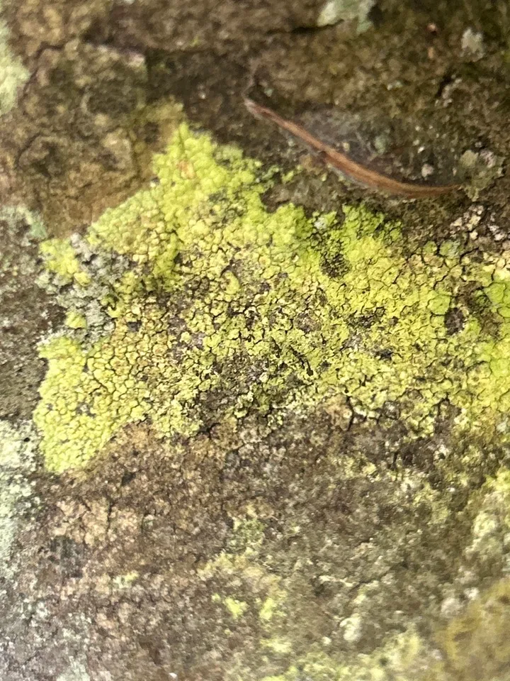
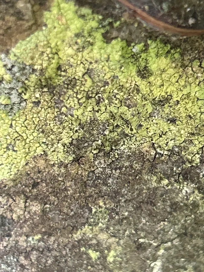
Dokumentasi Microcalicium arenarium menurut asosiasi foto pada record takson iNaturalist. Kredit: Fotografer tidak dicantumkan. Sumber observasi iNaturalist; pengunggah: Jonny Tostenson. Identitas mengikuti sumber; bukan penentuan spesimen independen.

Jenis
Fotografi
Kredit
Fotografer tidak dicantumkan. Sumber observasi iNaturalist; pengunggah: Jonny Tostenson
Lisensi
CC0 1.0
Sumber media
Buka entri sumber

Gambar menunjukkan objek menurut identifikasi sumber, bukan verifikasi independen spesimen oleh Fungi. Tidak ada skala baru yang ditambahkan.

Dokumentasi tambahan

06 / Rujukan

Sumber profil

  1. [1] Galloway, D. J. 1985: Flora of New Zealand: Lichens. Government Printer, Wellington. — Microcalicium arenariumBagian sumber: biota:82762755-cc7d-4cb0-8b36-4c4abd5608cb:3607e5f9-225c-4381-b579-6008f9732fd3
    Tanggal penghimpunan sumber: 2026-10-12
    Teks sumber: CC BY 4.0 · Galloway, D. J. 1985: Flora of New Zealand: Lichens. Government Printer, Wellington.
  2. [2] Catalogue of Life Base2026-09-11 — Microcalicium arenariumBagian sumber: ColDP NameUsage.tsv ID 42S6X; accepted species; source 2073
    Tanggal penghimpunan sumber: 2026-10-12
  3. [3] Dokumentasi visual — Microcalicium arenariumBagian sumber: Identitas mengikuti sumber dokumentasi; bukan penentuan spesimen independen.
    Tanggal penghimpunan sumber: 2026-10-17
    Media sumber: CC0 1.0 · Fotografer tidak dicantumkan. Sumber observasi iNaturalist; pengunggah: Jonny Tostenson
  4. [4] Dokumentasi visual — Microcalicium arenariumBagian sumber: Identitas mengikuti sumber dokumentasi; bukan penentuan spesimen independen.
    Tanggal penghimpunan sumber: 2026-10-17
    Teks sumber: CC0 1.0 · no rights reserved

Adaptasi teks: CC BY 4.0. Galloway, D. J. 1985: Flora of New Zealand: Lichens. Government Printer, Wellington. — Galloway, D. J. 1985: Flora of New Zealand: Lichens. Government Printer, Wellington. — Microcalicium arenarium

Uraian asli dan riwayat sumber

Riwayat atau halaman sumber

Ringkasan sumber

Growing on either pure crusts of or sometimes on mixed crusts of and a member of Cystococcaceae. In some areas mainly parasitic on. Apothecia very variable in size, 0.6-1.8(-2.5) mm high. Capitulum spherical, 0.14-0.28 mm diam. Mazaedium not protruding more than diam. of apothecia. Paraphyses not sclerotized, dissolving at an early stage. Excipulum in section reddish-brown, with a coarsely granular surface; smooth and pale greyish-brown in very long-stalked apothecia. Outer part of stalk covered by groups of sclerotized hyphae; inner part of stalk of pale and rather straight, interwoven hyphae.

Revisi sumber

Morfologi sumber

Growing on either pure crusts of or sometimes on mixed crusts of and a member of Cystococcaceae. In some areas mainly parasitic on. Apothecia very variable in size, 0.6-1.8(-2.5) mm high. Capitulum spherical, 0.14-0.28 mm diam. Mazaedium not protruding more than diam. of apothecia. Paraphyses not sclerotized, dissolving at an early stage. Excipulum in section reddish-brown, with a coarsely granular surface; smooth and pale greyish-brown in very long-stalked apothecia. Outer part of stalk covered by groups of sclerotized hyphae; inner part of stalk of pale and rather straight, interwoven hyphae. Asci when mature, broadly ellipsoid, 9-11 × 5-6 µm. Ascospores when mature 1-septate, 6.7-8.2 × 2.3-2.9 µm, distinctly ornamented with spirally arranged ridges. Chemistry: The aeruginose pigment of the apothecia and spores turns brown in K and reverts to intense green if excess acid is added. The structure of this pigment is not known.

Revisi sumber

Ekologi sumber

Growing on either pure crusts of or sometimes on mixed crusts of and a member of Cystococcaceae. In some areas mainly parasitic on. Apothecia very variable in size, 0.6-1.8(-2.5) mm high. Capitulum spherical, 0.14-0.28 mm diam. Mazaedium not protruding more than diam. of apothecia. Paraphyses not sclerotized, dissolving at an early stage. Excipulum in section reddish-brown, with a coarsely granular surface; smooth and pale greyish-brown in very long-stalked apothecia. Outer part of stalk covered by groups of sclerotized hyphae; inner part of stalk of pale and rather straight, interwoven hyphae. Asci when mature, broadly ellipsoid, 9-11 × 5-6 µm. Ascospores when mature 1-septate, 6.7-8.2 × 2.3-2.9 µm, distinctly ornamented with spirally arranged ridges. Chemistry: The aeruginose pigment of the apothecia and spores turns brown in K and reverts to intense green if excess acid is added. The structure of this pigment is not known.

Revisi sumber

Struktur reproduksi sumber

Growing on either pure crusts of or sometimes on mixed crusts of and a member of Cystococcaceae. In some areas mainly parasitic on. Apothecia very variable in size, 0.6-1.8(-2.5) mm high. Capitulum spherical, 0.14-0.28 mm diam. Mazaedium not protruding more than diam. of apothecia. Paraphyses not sclerotized, dissolving at an early stage. Excipulum in section reddish-brown, with a coarsely granular surface; smooth and pale greyish-brown in very long-stalked apothecia. Outer part of stalk covered by groups of sclerotized hyphae; inner part of stalk of pale and rather straight, interwoven hyphae. Asci when mature, broadly ellipsoid, 9-11 × 5-6 µm. Ascospores when mature 1-septate, 6.7-8.2 × 2.3-2.9 µm, distinctly ornamented with spirally arranged ridges.

Revisi sumber

Rujukan halaman ini

Fungi. Microcalicium arenarium. Rilis data fungi-2026-10-12-expansion-2. https://fungi-atlas.mgadib.chatgpt.site/jamur/microcalicium-arenarium/.

Status: impor sumber; pemeriksaan editorial per profil belum selesai. Untuk klaim biologis, gunakan rujukan pada bagian terkait dan periksa konteks sumber aslinya.