Ciri dan struktur
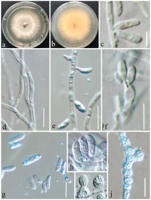
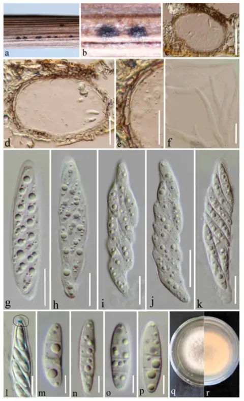
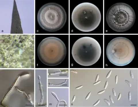
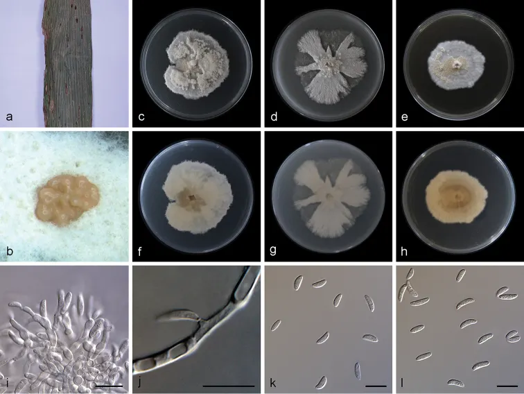
Characterised by conidiomata sporodochium-like and the size of conidia. Colonies on PDA attaining 48.0–61.5 mm in diameter after 15 days, felty, compact, erose or dentate, initially white, then becoming yellowish with age. Mycelium superficial, consisting of hyaline, smooth, branched, septate, 1.0–2.5 μm wide hyphae. Conidiomata sporodochium-like, appeared within 15 days or longer, formed in aerial mycelium or on agar surface, slimy, hyaline or orange, semi-submerged. Exudate occasionally appeared on old sporodochia. Reverse colorless to yellowish, due to the soluble pigment secreted. Conidiophores formed terminal or lateral with sympodial proliferation, solitary or aggregated. Most conidiophores tightly aggregated in a sporodochium, inconspicuous flat-tipped loci, irregularly branched, or borne directly on mycelial hyphae, straight or slightly curved, aseptate, guttulate, smooth-walled, apex subobtuse, base truncate. Conidiogenous cells of two types: some polyblastic, ampulliform, lageniform, with percurrent proliferations, 6.5–10.0 × 2.5–3.4 μm, neck up to 4.5 μm long, 1.0–1.5 μm wide, others straight or bent, smooth, cylindrical up to 10.0–11.5 μm long, 1.0–2.0 μm wide. Conidia aseptate, mostly lunate, a few ellipsoid and cylindrical, hyaline, straight or curved, obtuse, 0–2 guttulate in mature conidia, 6.8–10.0 × 2.4–3.5 μm. Chlamydospores was not observed. Sexual morph: unknown. Colonies on OA 58.1–61.5 mm in diameter after 15 days, entire, flat, white, lobate and radially margin, aerial mycelia cottony or sparse. Less exudate. Reverse white. Vegetative hyphae hyaline, abundant, branched, septate, thin-walled.
Habitat dan peran ekologis
Colonies on PDA attaining 48.0–61.5 mm in diameter after 15 days, felty, compact, erose or dentate, initially white, then becoming yellowish with age. Mycelium superficial, consisting of hyaline, smooth, branched, septate, 1.0–2.5 μm wide hyphae. Conidiomata sporodochium-like, appeared within 15 days or longer, formed in aerial mycelium or on agar surface, slimy, hyaline or orange, semi-submerged. Exudate occasionally appeared on old sporodochia. Reverse colorless to yellowish, due to the soluble pigment secreted. Conidiophores formed terminal or lateral with sympodial proliferation, solitary or aggregated. Most conidiophores tightly aggregated in a sporodochium, inconspicuous flat-tipped loci, irregularly branched, or borne directly on mycelial hyphae, straight or slightly curved, aseptate, guttulate, smooth-walled, apex subobtuse, base truncate. Conidiogenous cells of two types: some polyblastic, ampulliform, lageniform, with percurrent proliferations, 6.5–10.0 × 2.5–3.4 μm, neck up to 4.5 μm long, 1.0–1.5 μm wide, others straight or bent, smooth, cylindrical up to 10.0–11.5 μm long, 1.0–2.0 μm wide. Conidia aseptate, mostly lunate, a few ellipsoid and cylindrical, hyaline, straight or curved, obtuse, 0–2 guttulate in mature conidia, 6.8–10.0 × 2.4–3.5 μm.
- Habitat atau konteks temuan
- kultur yang didokumentasikan
- Substrat
- Informasi belum tersedia pada sumber rilis ini.
- Peran atau konteks
- Belum dikategorikan pada rilis ini.
Reproduksi
Characterised by conidiomata sporodochium-like and the size of conidia. Colonies on PDA attaining 48.0–61.5 mm in diameter after 15 days, felty, compact, erose or dentate, initially white, then becoming yellowish with age. Mycelium superficial, consisting of hyaline, smooth, branched, septate, 1.0–2.5 μm wide hyphae. Conidiomata sporodochium-like, appeared within 15 days or longer, formed in aerial mycelium or on agar surface, slimy, hyaline or orange, semi-submerged. Exudate occasionally appeared on old sporodochia. Reverse colorless to yellowish, due to the soluble pigment secreted. Conidiophores formed terminal or lateral with sympodial proliferation, solitary or aggregated. Most conidiophores tightly aggregated in a sporodochium, inconspicuous flat-tipped loci, irregularly branched, or borne directly on mycelial hyphae, straight or slightly curved, aseptate, guttulate, smooth-walled, apex subobtuse, base truncate.
Catatan identitas dan cakupan
- Nama diterima mengikuti Catalogue of Life Base2026-09-11; uraian mempertahankan konsep dan nama sumber aslinya.
- Preserve original treatment localities, hosts and substrate scope. Materials examined describe documented specimens, not the full global range.
- Impor dan terjemahan otomatis belum diperiksa editorial per profil. Struktur reproduktif yang disebutkan tidak membuktikan seluruh siklus hidup diketahui.
- Pengukuran pada uraian mengikuti spesimen, kultur, dan metode sumber; angka tidak diperluas ke semua populasi.
Catatan distribusi dalam narasi mengikuti sumbernya. Dokumentasi spesimen atau foto mengikuti catatan sumber. Bukti wilayah Indonesia dan penilaian konservasi tidak disimpulkan dari gambar.
Melihat dengan konteks
Microdochium yunnanense (SAUCC1011) a leaves of host plant b sporodochia on media surface c–e surface of colony after 15 days on PDA (c), OA (d), MEA (e) f–h reverse of colony after 15 days on PDA (f), OA (g), MEA (h) i sporodochial conidiophores and conidiogenous cells with conidia j conidiogenous cells k–l conidia. Scale bars: 10μm (i–l).
- Jenis
- Pelat ilmiah
- Kredit
- Shengting Huang, Jiwen Xia, Xiuguo Zhang, Wenxiu Sun, Zhuang Li
- Lisensi
- CC BY 4.0
- Sumber media
- Buka entri sumber
Sumber profil
- [1] Two new species of Microdochium from Indocalamus longiauritus in south-western China — Microdochium yunnanense
- [2] Catalogue of Life Base2026-09-11 — Microdochium yunnanense
- [3] Dokumentasi visual — Microdochium yunnanense
Rujukan halaman ini
Fungi. Microdochium yunnanense. Rilis data fungi-2026-10-12-expansion-2. https://fungi-atlas.mgadib.chatgpt.site/jamur/microdochium-yunnanense/.
Status: impor sumber; pemeriksaan editorial per profil belum selesai. Untuk klaim biologis, gunakan rujukan pada bagian terkait dan periksa konteks sumber aslinya.