Ciri dan struktur
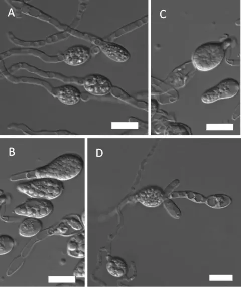
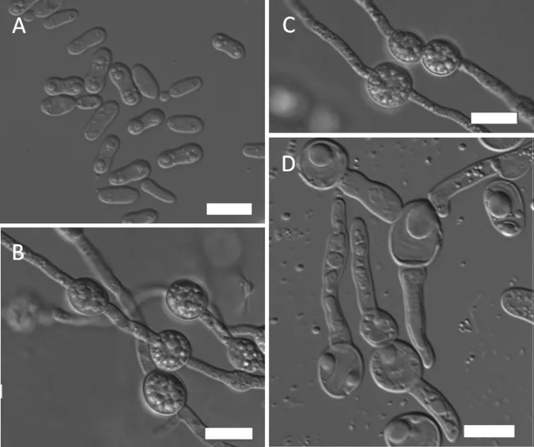
The physiological profiles of the three strains of the novel species were almost identical. In YM broth after 3 days at 15 °C, the cells are ovoid to elongated (3.5–7 × 3.5–5 µm) and proliferate by polar budding (Fig. 2A). Streak culture for 1 week at 15 °C on YM agar produces colonies that are white to yellowish-cream, round, convex and smooth with an entire margin. After 2 weeks in Dalmau plate culture on CM agar at 15 °C, pseudohyphae and true hyphae are formed. Teliospores were observed after 2 months at 10 °C terminally and intercalarily on the hyphae on CM agar (Fig. 2B). Teliospores are spherical, 8–13 µm in diameter, single and in short chains of 2–4 spores (Fig. 2C). Teliospores may germinate by a bud-like projection (Fig. 2D). The fermentation of sugars is absent. Glucose, galactose, l-sorbose, d-ribose, d-xylose, l-arabinose, l-rhamnose, sucrose, maltose, trehalose, methyl α-d-glucoside, cellobiose, salicin, arbutin, melibiose, lactose, raffinose, melezitose, inulin, glycerol, erythritol, ribitol, xylitol, l-arabinitol, d-glucitol, d-mannitol, galactitol, 2-keto-d-gluconate, d-gluconate, d-glucuronate, succinate, citrate and ethanol are assimilated. No growth occurs in d-glucosamine, d-arabinose, myo -inositol, d-glucono-1, 5-lactone, 5-keto-d-gluconate, dl-lactate or methanol. For the assimilation of nitrogen compounds, growth on nitrate, nitrite, l-lysine, glucosamine and d-tryptophan is positive, whereas on ethylamine, cadaverine, creatine, creatinine or imidazole, it is negative. There is no growth in the presence of 5% glucose medium with 10% NaCl and 0.01% cycloheximide.
Habitat dan peran ekologis
holotype, NYNU 18562 culture was isolated from the soil of the Lianhuashan National Forest Park, Jilin Province, China, in September 2018. The holotype culture is preserved in a metabolically inactive state at Microbiology Lab, Nanyang Normal University, Henan, China. Ex-type cultures are deposited at the China Centre of Industrial Culture Collection (CICC), Beijing, China, as CICC 33355, and at the CBS Yeast Collection of the Westerdijk Fungal Biodiversity Institute, Utrecht, the Netherlands, as CBS 15868.
- Habitat atau konteks temuan
- hutan; kultur yang didokumentasikan
- Substrat
- Informasi belum tersedia pada sumber rilis ini.
- Peran atau konteks
- Belum dikategorikan pada rilis ini.
Reproduksi
The physiological profiles of the three strains of the novel species were almost identical. In YM broth after 3 days at 15 °C, the cells are ovoid to elongated (3.5–7 × 3.5–5 µm) and proliferate by polar budding (Fig. 2A). Streak culture for 1 week at 15 °C on YM agar produces colonies that are white to yellowish-cream, round, convex and smooth with an entire margin. After 2 weeks in Dalmau plate culture on CM agar at 15 °C, pseudohyphae and true hyphae are formed. Teliospores were observed after 2 months at 10 °C terminally and intercalarily on the hyphae on CM agar (Fig. 2B). Teliospores are spherical, 8–13 µm in diameter, single and in short chains of 2–4 spores (Fig. 2C). Teliospores may germinate by a bud-like projection (Fig. 2D).
Catatan identitas dan cakupan
- Nama diterima mengikuti Catalogue of Life Base2026-09-11; uraian mempertahankan konsep dan nama sumber aslinya.
- Preserve original treatment localities, hosts and substrate scope. Materials examined describe documented specimens, not the full global range.
- Impor dan terjemahan otomatis belum diperiksa editorial per profil. Struktur reproduktif yang disebutkan tidak membuktikan seluruh siklus hidup diketahui.
- Pengukuran pada uraian mengikuti spesimen, kultur, dan metode sumber; angka tidak diperluas ke semua populasi.
Catatan distribusi dalam narasi mengikuti sumbernya. Dokumentasi spesimen atau foto mengikuti catatan sumber. Bukti wilayah Indonesia dan penilaian konservasi tidak disimpulkan dari gambar.
Melihat dengan konteks
Morphology of Mrakia panshiensis (NYNU 18562 T) A budding cells B true hyphae with clamp connections and teliospores C teliospores in pairs D teliospores produced with a bud-like projection. Scale bars: 10 μm.
- Jenis
- Pelat ilmiah
- Kredit
- Kai-Hong Zhang, Cheng-Feng Shi, Chun-Yue Chai, Feng-Li Hui
- Lisensi
- CC BY 4.0
- Sumber media
- Buka entri sumber
Sumber profil
- [1] Mrakia panshiensis sp. nov. a new member of the Cystofilobasidiales from soil in China, and description of the teleomorphic-stage of M. arctica — Mrakia panshiensis
- [2] Catalogue of Life Base2026-09-11 — Mrakia panshiensis
- [3] Dokumentasi visual — Mrakia panshiensis
Rujukan halaman ini
Fungi. Mrakia panshiensis. Rilis data fungi-2026-10-12-expansion-2. https://fungi-atlas.mgadib.chatgpt.site/jamur/mrakia-panshiensis/.
Status: impor sumber; pemeriksaan editorial per profil belum selesai. Untuk klaim biologis, gunakan rujukan pada bagian terkait dan periksa konteks sumber aslinya.